 Movie
Movie Controller


+ Open data

- Basic information
| Entry | Database: PDB / ID: 8afy | ||||||
|---|---|---|---|---|---|---|---|
| Title | Subtomogram average of membrane-bound Atg18 oligomers | ||||||
 Components Components | Autophagy-related protein 18 | ||||||
 Keywords Keywords |  MEMBRANE PROTEIN / autophagy / membrane remodeling / PIP binding / PI3P / PI(3 / 5)P2 / lipid binding protein MEMBRANE PROTEIN / autophagy / membrane remodeling / PIP binding / PI3P / PI(3 / 5)P2 / lipid binding protein | ||||||
| Function / homology |  Function and homology information Function and homology informationregulation of phosphatidylinositol biosynthetic process / PAS complex / 1-phosphatidyl-1D-myo-inositol 3,5-bisphosphate metabolic process / phagophore / vacuolar protein processing / positive regulation of vacuole organization / Macroautophagy / cytoplasm to vacuole targeting by the Cvt pathway / nucleophagy / pexophagy ...regulation of phosphatidylinositol biosynthetic process / PAS complex / 1-phosphatidyl-1D-myo-inositol 3,5-bisphosphate metabolic process / phagophore / vacuolar protein processing / positive regulation of vacuole organization / Macroautophagy / cytoplasm to vacuole targeting by the Cvt pathway / nucleophagy / pexophagy / protein localization to phagophore assembly site / piecemeal microautophagy of the nucleus / phagophore assembly site membrane / late endosome to vacuole transport / phosphatidylinositol-3-phosphate binding / fungal-type vacuole membrane / phagophore assembly site / phosphatidylinositol-4-phosphate binding / phosphatidylinositol-3,5-bisphosphate binding / vacuolar membrane / extrinsic component of membrane / autophagosome assembly / ubiquitin binding / cell periphery / macroautophagy / protein transport / endosome membrane / endosome / protein-containing complex / cytosolSimilarity search - Function | ||||||
| Biological species |  Saccharomyces cerevisiae (brewer's yeast) Saccharomyces cerevisiae (brewer's yeast) | ||||||
| Method | ELECTRON MICROSCOPY / subtomogram averaging / cryo EM / Resolution: 26 Å | ||||||
 Authors Authors | Mann, D. / Fromm, S. / Martinez-Sanchez, A. / Gopaldass, N. / Mayer, A. / Sachse, C. | ||||||
| Funding support |  Germany, 1items Germany, 1items
| ||||||
 Citation Citation | Journal: To Be Published Title: Cryo-EM structures of Atg18 oligomers reveal a tilted structural scaffold for Atg2 at the isolation membrane Authors: Mann, D. / Fromm, S.A. / Martinez-Sanchez, A. / Gopaldass, N. / Mayer, A. / Sachse, C. | ||||||
| History |
|
- Structure visualization
| Structure viewer | Molecule: MolmilJmol/JSmol |
|---|
- Downloads & links
-Download
| PDBx/mmCIF format | 8afy.cif.gz | 610.7 KB | Display | PDBx/mmCIF format |
|---|---|---|---|---|
| PDB format | pdb8afy.ent.gz | 475.3 KB | Display | PDB format |
| PDBx/mmJSON format | 8afy.json.gz | Tree view | PDBx/mmJSON format | |
| Others |  Other downloads Other downloads |
-Validation report
| Arichive directory | https://data.pdbj.org/pub/pdb/validation_reports/af/8afyftp://data.pdbj.org/pub/pdb/validation_reports/af/8afy | HTTPS FTP |
|---|
-Related structure data
| Related structure data |  15412MC  8afqC  8afwC 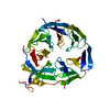 8afxC M: map data used to model this data C: citing same article ( |
|---|---|
| Similar structure data |


-Links
 PDBj
PDBj
- Assembly
| Deposited unit | 
|
|---|---|
| 1 |
|
-Components
| #1: Protein | Mass: 55045.777 Da / Num. of mol.: 8 Source method: isolated from a genetically manipulated source Source: (gene. exp.) Saccharomyces cerevisiae (brewer's yeast)Strain: ATCC 204508 / S288c / Gene: ATG18, AUT10, CVT18, NMR1, SVP1, YFR021W / Production host:  Escherichia coli (E. coli) / References: UniProt: P43601 Escherichia coli (E. coli) / References: UniProt: P43601 |
|---|
-Experimental details
-Experiment
| Experiment | Method: ELECTRON MICROSCOPY |
|---|---|
| EM experiment | Aggregation state: PARTICLE / 3D reconstruction method: subtomogram averaging |
- Sample preparation
| Component | Name: Subtomogram average of membrane-bound Atg18 / Type: COMPLEX / Entity ID: all / Source: RECOMBINANT |
|---|---|
| Molecular weight | Experimental value: NO |
| Source (natural) | Organism: Saccharomyces cerevisiae (brewer's yeast) |
| Source (recombinant) | Organism: Escherichia coli (E. coli) |
| Buffer solution | pH: 7.2 |
| Specimen | Embedding applied: NO / Shadowing applied: NO / Staining applied: NO / Vitrification applied: YES |
| Vitrification | Instrument: LEICA EM GP / Cryogen name: ETHANE / Humidity: 99 % / Chamber temperature: 291 K |
- Electron microscopy imaging
| Experimental equipment |  Model: Titan Krios / Image courtesy: FEI Company |
|---|---|
| Microscopy | Model: TFS KRIOS |
| Electron gun | Electron source: FIELD EMISSION GUN / Accelerating voltage: 300 kV / Illumination mode: FLOOD BEAM |
| Electron lens | Mode: BRIGHT FIELDBright-field microscopy / Nominal defocus max: 0 nm / Nominal defocus min: 0 nm / Alignment procedure: COMA FREE |
| Specimen holder | Cryogen: NITROGEN / Specimen holder model: FEI TITAN KRIOS AUTOGRID HOLDER |
| Image recording | Electron dose: 3 e/Å2 / Avg electron dose per subtomogram: 120 e/Å2 / Detector mode: COUNTING / Film or detector model: GATAN K2 SUMMIT (4k x 4k) |
| EM imaging optics | Phase plate: VOLTA PHASE PLATE |
- Processing
| EM software |
| |||||||||||||||||||||
|---|---|---|---|---|---|---|---|---|---|---|---|---|---|---|---|---|---|---|---|---|---|---|
| CTF correction | Type: NONE | |||||||||||||||||||||
| Symmetry | Point symmetry: C1 (asymmetric) | |||||||||||||||||||||
| 3D reconstruction | Resolution: 26 Å / Resolution method: OTHER / Num. of particles: 8300 / Algorithm: BACK PROJECTION / Details: Mask-less FDR-FSC at 1% FDR / Symmetry type: POINT | |||||||||||||||||||||
| EM volume selection | Num. of tomograms: 16 / Num. of volumes extracted: 8300 | |||||||||||||||||||||
| Atomic model building | Protocol: RIGID BODY FIT / Space: REAL Details: Fitted 4x dimer "T" from Atg18 filament structure to interpret the density as a tetramer of dimers |
