 Movie
Movie Controller


+ Open data

- Basic information
| Entry |  | |||||||||
|---|---|---|---|---|---|---|---|---|---|---|
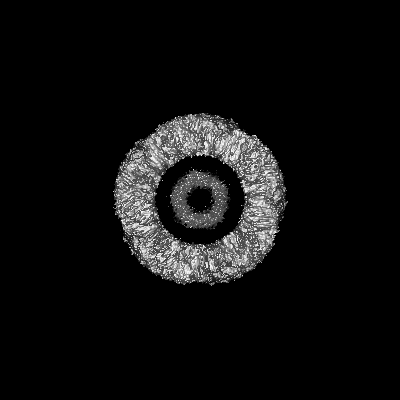
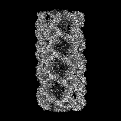
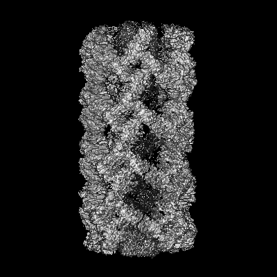
| Title | Tube assembly of Atg18-WT | |||||||||
 Map data Map data | sharp map from CryoSPARC software | |||||||||
 Sample Sample |
| |||||||||
 Keywords Keywords |  autophagy / membrane remodeling / PIP binding / PI3P / PI(3 / 5)P2 / lipid binding protein / MEMBRANE PROTEIN autophagy / membrane remodeling / PIP binding / PI3P / PI(3 / 5)P2 / lipid binding protein / MEMBRANE PROTEIN | |||||||||
| Function / homology |  Function and homology information Function and homology informationregulation of phosphatidylinositol biosynthetic process / PAS complex / 1-phosphatidyl-1D-myo-inositol 3,5-bisphosphate metabolic process / phagophore / vacuolar protein processing / positive regulation of vacuole organization / Macroautophagy / cytoplasm to vacuole targeting by the Cvt pathway / nucleophagy / pexophagy ...regulation of phosphatidylinositol biosynthetic process / PAS complex / 1-phosphatidyl-1D-myo-inositol 3,5-bisphosphate metabolic process / phagophore / vacuolar protein processing / positive regulation of vacuole organization / Macroautophagy / cytoplasm to vacuole targeting by the Cvt pathway / nucleophagy / pexophagy / protein localization to phagophore assembly site / piecemeal microautophagy of the nucleus / phagophore assembly site membrane / late endosome to vacuole transport / phosphatidylinositol-3-phosphate binding / fungal-type vacuole membrane / phagophore assembly site / phosphatidylinositol-4-phosphate binding / phosphatidylinositol-3,5-bisphosphate binding / vacuolar membrane / extrinsic component of membrane / autophagosome assembly / ubiquitin binding / cell periphery / macroautophagy / protein transport / endosome membrane / endosome / protein-containing complex / cytosolSimilarity search - Function | |||||||||
| Biological species |  Saccharomyces cerevisiae (brewer's yeast) Saccharomyces cerevisiae (brewer's yeast) | |||||||||
| Method | helical reconstruction / cryo EM / Resolution: 3.8 Å | |||||||||
 Authors Authors | Mann D / Fromm S / Martinez-Sanchez A / Gopaldass N / Mayer A / Sachse C | |||||||||
| Funding support |  Germany, 1 items Germany, 1 items
| |||||||||
 Citation Citation | Journal: To Be Published Title: Cryo-EM structures of Atg18 oligomers reveal a tilted structural scaffold for Atg2 at the isolation membrane Authors: Mann D / Fromm SA / Martinez-Sanchez A / Gopaldass N / Mayer A / Sachse C | |||||||||
| History |
|
- Structure visualization
| Supplemental images |
|---|

- Downloads & links
-EMDB archive
| Map data | emd_15410.map.gz | 229.1 MB | EMDB map data format | |
|---|---|---|---|---|
| Header (meta data) | emd-15410-v30.xmlemd-15410.xml | 15.1 KB 15.1 KB | Display Display | EMDB header |
| FSC (resolution estimation) | emd_15410_fsc.xml | 13.2 KB | Display | FSC data file |
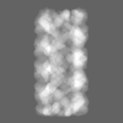
| Images | 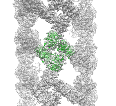 emd_15410.png emd_15410.png | 171 KB | ||
| Masks | emd_15410_msk_1.map | 244.1 MB | Mask map | |
| Filedesc metadata | emd-15410.cif.gz | 5.6 KB | ||
| Others | emd_15410_half_map_1.map.gzemd_15410_half_map_2.map.gz | 225.9 MB 225.9 MB | ||
| Archive directory |  http://ftp.pdbj.org/pub/emdb/structures/EMD-15410ftp://ftp.pdbj.org/pub/emdb/structures/EMD-15410 http://ftp.pdbj.org/pub/emdb/structures/EMD-15410ftp://ftp.pdbj.org/pub/emdb/structures/EMD-15410 | HTTPS FTP |
-Related structure data
| Related structure data |  8afwMC  8afqC  8afxC  8afyC M: atomic model generated by this map C: citing same article ( |
|---|---|
| Similar structure data |


-Links
| EMDB pages | EMDB (EBI/PDBe) / EMDataResource |
|---|---|
| Related items in Molecule of the Month |

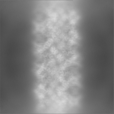
-Map
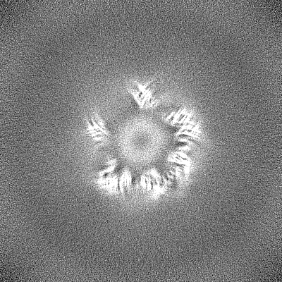
| File | Download / File: emd_15410.map.gz / Format: CCP4 / Size: 244.1 MB / Type: IMAGE STORED AS FLOATING POINT NUMBER (4 BYTES) | ||||||||||||||||||||||||||||||||||||
|---|---|---|---|---|---|---|---|---|---|---|---|---|---|---|---|---|---|---|---|---|---|---|---|---|---|---|---|---|---|---|---|---|---|---|---|---|---|
| Annotation | sharp map from CryoSPARC software | ||||||||||||||||||||||||||||||||||||
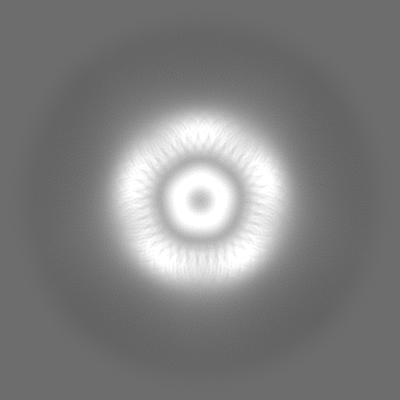
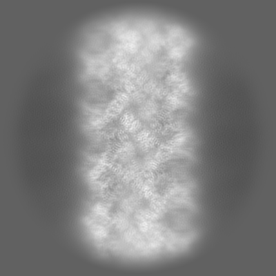
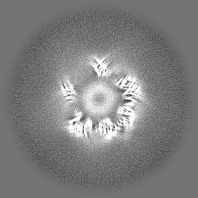
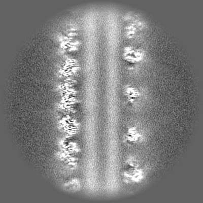
| Projections & slices | Image control
Images are generated by Spider. | ||||||||||||||||||||||||||||||||||||
| Voxel size | X=Y=Z: 1.35 Å | ||||||||||||||||||||||||||||||||||||
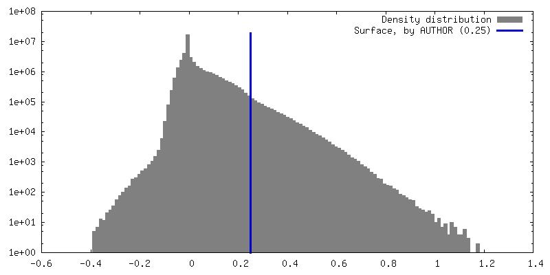
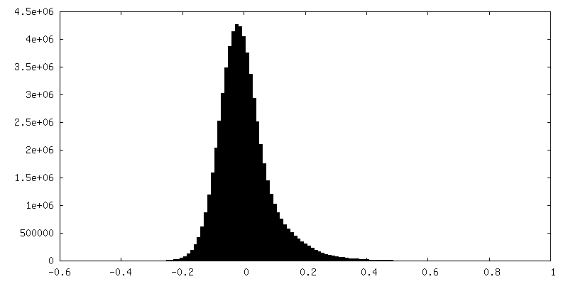
| Density |
| ||||||||||||||||||||||||||||||||||||
| Symmetry | Space group: 1 | ||||||||||||||||||||||||||||||||||||
| Details | EMDB XML:
|
 Z (Sec.)
 Y (Row.)
 X (Col.)


-Supplemental data
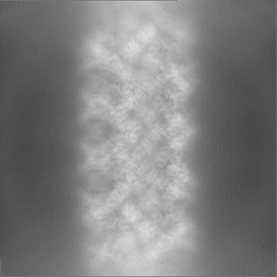
-Mask #1
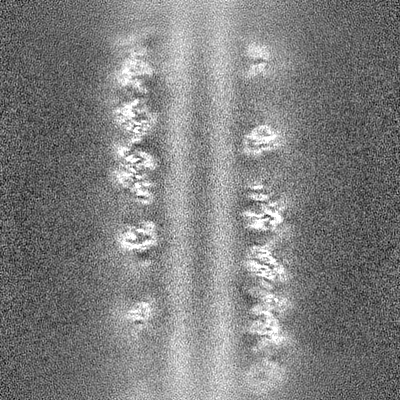
| File | emd_15410_msk_1.map | ||||||||||||
|---|---|---|---|---|---|---|---|---|---|---|---|---|---|
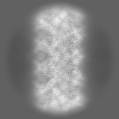
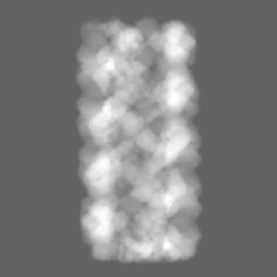
| Projections & Slices |
| ||||||||||||
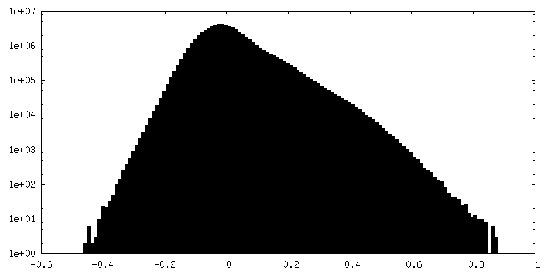
| Density Histograms |


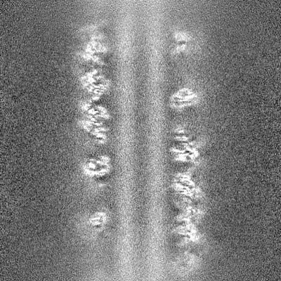
-Half map: half map A
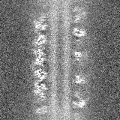
| File | emd_15410_half_map_1.map | ||||||||||||
|---|---|---|---|---|---|---|---|---|---|---|---|---|---|
| Annotation | half map A | ||||||||||||
| Projections & Slices |
| ||||||||||||
| Density Histograms |


-Half map: half map B
| File | emd_15410_half_map_2.map | ||||||||||||
|---|---|---|---|---|---|---|---|---|---|---|---|---|---|
| Annotation | half map B | ||||||||||||
| Projections & Slices |
| ||||||||||||
| Density Histograms |


- Sample components
-Entire : Filament assembly of Atg18-WT
| Entire | Name: Filament assembly of Atg18-WT |
|---|---|
| Components |
|
-Supramolecule #1: Filament assembly of Atg18-WT
| Supramolecule | Name: Filament assembly of Atg18-WT / type: complex / ID: 1 / Parent: 0 / Macromolecule list: all |
|---|---|
| Source (natural) | Organism: Saccharomyces cerevisiae (brewer's yeast) |
-Macromolecule #1: Autophagy-related protein 18
| Macromolecule | Name: Autophagy-related protein 18 / type: protein_or_peptide / ID: 1 / Number of copies: 4 / Enantiomer: LEVO |
|---|---|
| Source (natural) | Organism: Saccharomyces cerevisiae (brewer's yeast) / Strain: ATCC 204508 / S288c |
| Molecular weight | Theoretical: 55.15793 KDa |
| Recombinant expression | Organism:  Escherichia coli (E. coli) Escherichia coli (E. coli) |
| Sequence | String: MSDSSPTINF INFNQTGTCI SLGTSKGFKI FNCEPFGKFY SEDSGGYAIV EMLFSTSLLA LVGIGDQPAL SPRRLRIINT KKHSIICEV TFPTSILSVK MNKSRLVVLL QEQIYIYDIN TMRLLHTIET NPNPRGLMAM SPSVANSYLV YPSPPKVINS E IKAHATTN ...String: MSDSSPTINF INFNQTGTCI SLGTSKGFKI FNCEPFGKFY SEDSGGYAIV EMLFSTSLLA LVGIGDQPAL SPRRLRIINT KKHSIICEV TFPTSILSVK MNKSRLVVLL QEQIYIYDIN TMRLLHTIET NPNPRGLMAM SPSVANSYLV YPSPPKVINS E IKAHATTN NITLSVGGNT ETSFKRDQQD AGHSDISDLD QYSSFTKRDD ADPTSSNGGN SSIIKNGDVI VFNLETLQPT MV IEAHKGE IAAMAISFDG TLMATASDKG TIIRVFDIET GDKIYQFRRG TYATRIYSIS FSEDSQYLAV TGSSKTVHIF KLG HSMSNN KLDSDDSNME EAAADDSSLD TTSIDALSDE ENPTRLAREP YVDASRKTMG RMIRYSSQKL SRRAARTLGQ IFPI KVTSL LESSRHFASL KLPVETNSHV MTISSIGSPI DIDTSEYPEL FETGNSASTE SYHEPVMKMV PIRVVSSDGY LYNFV MDPE RGGDCLILSQ YSILMD UniProtKB: Autophagy-related protein 18 |
-Experimental details
-Structure determination
| Method | cryo EM |
|---|---|
 Processing Processing | helical reconstruction |
| Aggregation state | filament |
-Sample preparation
| Buffer | pH: 7.2 |
|---|---|
| Vitrification | Cryogen name: ETHANE / Chamber humidity: 99 % / Chamber temperature: 291 K / Instrument: LEICA EM GP |
- Electron microscopy
| Microscope | TFS KRIOS |
|---|---|
| Electron beam | Acceleration voltage: 300 kV / Electron source: FIELD EMISSION GUN |
| Electron optics | Illumination mode: FLOOD BEAM / Imaging mode: BRIGHT FIELDBright-field microscopy / Nominal defocus max: 3.0 µm / Nominal defocus min: 1.0 µm |
| Sample stage | Specimen holder model: FEI TITAN KRIOS AUTOGRID HOLDER / Cooling holder cryogen: NITROGEN |
| Image recording | Film or detector model: GATAN K2 SUMMIT (4k x 4k) / Detector mode: COUNTING / Number real images: 1812 / Average electron dose: 90.0 e/Å2 |
| Experimental equipment |  Model: Titan Krios / Image courtesy: FEI Company |
-Image processing
| Startup model | Type of model: NONE |
|---|---|
| Final angle assignment | Type: NOT APPLICABLE / Software - Name: cryoSPARC |
| Final reconstruction | Applied symmetry - Helical parameters - Δz: 19.4 Å Applied symmetry - Helical parameters - Δ&Phi: 70 ° Applied symmetry - Helical parameters - Axial symmetry: C1 (asymmetric) Resolution.type: BY AUTHOR / Resolution: 3.8 Å / Resolution method: FSC 0.143 CUT-OFF / Software - Name: cryoSPARC / Number images used: 133981 |
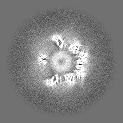
| FSC plot (resolution estimation) | 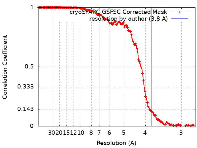 |
-Atomic model buiding 1
| Details | Initial PDB was derived from the Atg18-PR72AA filament structure, subunits were individually docked inside the density and the PR72AA loop was manually modified in Coot |
|---|---|
| Refinement | Space: REAL / Protocol: RIGID BODY FIT / Target criteria: Correlation coefficient |
| Output model | PDB-8afw: |
