 Movie
Movie Controller

[English] 日本語
 Yorodumi
- PDB-7yni: Structure of human SGLT1-MAP17 complex bound with substrate 4D4FD... -
+ Open data

- Basic information
| Entry | Database: PDB / ID: 7yni | ||||||||||||
|---|---|---|---|---|---|---|---|---|---|---|---|---|---|
| Title | Structure of human SGLT1-MAP17 complex bound with substrate 4D4FDG in the occluded conformation | ||||||||||||
 Components Components |
| ||||||||||||
 Keywords Keywords |  PROTEIN TRANSPORT / glucose transporter / SGLT / sodium glucose transporter / membrane protein PROTEIN TRANSPORT / glucose transporter / SGLT / sodium glucose transporter / membrane protein | ||||||||||||
| Function / homology |  Function and homology information Function and homology informationmyo-inositol:sodium symporter activity / pentose transmembrane transporter activity / fucose transmembrane transporter activity / galactose:sodium symporter activity / pentose transmembrane transport / myo-inositol transport / intestinal hexose absorption / Defective SLC5A1 causes congenital glucose/galactose malabsorption (GGM) / Intestinal hexose absorption / fucose transmembrane transport ...myo-inositol:sodium symporter activity / pentose transmembrane transporter activity / fucose transmembrane transporter activity / galactose:sodium symporter activity / pentose transmembrane transport / myo-inositol transport / intestinal hexose absorption / Defective SLC5A1 causes congenital glucose/galactose malabsorption (GGM) / Intestinal hexose absorption / fucose transmembrane transport / intestinal D-glucose absorption / galactose transmembrane transporter activity / alpha-glucoside transport / glucose:sodium symporter activity / alpha-glucoside transmembrane transporter activity / galactose transmembrane transport / glucose transmembrane transporter activity / water transmembrane transporter activity / D-glucose transmembrane transporter activity / Cellular hexose transport / renal glucose absorption / glucose import across plasma membrane / glucose transmembrane transport / transepithelial water transport / intracellular organelle / sodium ion import across plasma membrane / intracellular vesicle / sodium ion transport / transport across blood-brain barrier / : / brush border membrane / early endosome / apical plasma membrane / perinuclear region of cytoplasm / extracellular exosome / membrane / plasma membraneSimilarity search - Function | ||||||||||||
| Biological species |  Homo sapiens (human) Homo sapiens (human) | ||||||||||||
| Method | ELECTRON MICROSCOPY / single particle reconstruction / cryo EM / Resolution: 3.26 Å | ||||||||||||
 Authors Authors | Chen, L. / Niu, Y. / Cui, W. | ||||||||||||
| Funding support |  China, 3items China, 3items
| ||||||||||||
 Citation Citation | Journal: Nat Commun / Year: 2023 Title: Structures of human SGLT in the occluded state reveal conformational changes during sugar transport. Authors: Wenhao Cui / Yange Niu / Zejian Sun / Rui Liu / Lei Chen / Abstract: Sodium-Glucose Cotransporters (SGLT) mediate the uphill uptake of extracellular sugars and play fundamental roles in sugar metabolism. Although their structures in inward-open and outward-open ...Sodium-Glucose Cotransporters (SGLT) mediate the uphill uptake of extracellular sugars and play fundamental roles in sugar metabolism. Although their structures in inward-open and outward-open conformations are emerging from structural studies, the trajectory of how SGLTs transit from the outward-facing to the inward-facing conformation remains unknown. Here, we present the cryo-EM structures of human SGLT1 and SGLT2 in the substrate-bound state. Both structures show an occluded conformation, with not only the extracellular gate but also the intracellular gate tightly sealed. The sugar substrate are caged inside a cavity surrounded by TM1, TM2, TM3, TM6, TM7, and TM10. Further structural analysis reveals the conformational changes associated with the binding and release of substrates. These structures fill a gap in our understanding of the structural mechanisms of SGLT transporters. | ||||||||||||
| History |
|
- Structure visualization
| Structure viewer | Molecule: MolmilJmol/JSmol |
|---|
- Downloads & links
-Download
| PDBx/mmCIF format | 7yni.cif.gz | 126 KB | Display | PDBx/mmCIF format |
|---|---|---|---|---|
| PDB format | pdb7yni.ent.gz | 90.8 KB | Display | PDB format |
| PDBx/mmJSON format | 7yni.json.gz | Tree view | PDBx/mmJSON format | |
| Others |  Other downloads Other downloads |
-Validation report
| Arichive directory | https://data.pdbj.org/pub/pdb/validation_reports/yn/7yniftp://data.pdbj.org/pub/pdb/validation_reports/yn/7yni | HTTPS FTP |
|---|
-Related structure data
| Related structure data | 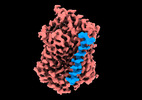 33962MC 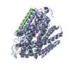 7ynjC 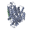 7ynkC M: map data used to model this data C: citing same article ( |
|---|---|
| Similar structure data |
-Links
 PDBj
PDBj

- Assembly
| Deposited unit | 
|
|---|---|
| 1 |
|
-Components
| #1: Protein | / Na(+)/glucose cotransporter 1 / High affinity sodium-glucose cotransporter / Solute carrier family 5 member 1 Mass: 73557.703 Da / Num. of mol.: 1 Source method: isolated from a genetically manipulated source Source: (gene. exp.) Homo sapiens (human) / Gene: SLC5A1, NAGT, SGLT1 / Production host: Homo sapiens (human) / References: UniProt: P13866 |
|---|---|
| #2: Protein | Mass: 12235.000 Da / Num. of mol.: 1 Source method: isolated from a genetically manipulated source Source: (gene. exp.) Homo sapiens (human) / Gene: PDZK1IP1, MAP17 / Production host: Homo sapiens (human) / References: UniProt: Q13113 |
| #3: Sugar | ChemComp-KQC / (  Type: D-saccharide, beta linking / Mass: 182.147 Da / Num. of mol.: 1 / Source method: obtained synthetically / Formula: C6H11FO5 / Feature type: SUBJECT OF INVESTIGATION Type: D-saccharide, beta linking / Mass: 182.147 Da / Num. of mol.: 1 / Source method: obtained synthetically / Formula: C6H11FO5 / Feature type: SUBJECT OF INVESTIGATION |
| Has ligand of interest | Y |
-Experimental details
-Experiment
| Experiment | Method: ELECTRON MICROSCOPY |
|---|---|
| EM experiment | Aggregation state: PARTICLE / 3D reconstruction method: single particle reconstruction |
- Sample preparation
| Component | Name: human SGLT1-MAP17 complex / Type: COMPLEX / Entity ID: #1-#2 / Source: RECOMBINANT |
|---|---|
| Source (natural) | Organism: Homo sapiens (human) |
| Source (recombinant) | Organism: Homo sapiens (human) |
| Buffer solution | pH: 7.5 |
| Specimen | Embedding applied: NO / Shadowing applied: NO / Staining applied: NO / Vitrification applied: YES |
| Vitrification | Cryogen name: ETHANE |
- Electron microscopy imaging
| Experimental equipment |  Model: Titan Krios / Image courtesy: FEI Company |
|---|---|
| Microscopy | Model: FEI TITAN KRIOS |
| Electron gun | Electron source: FIELD EMISSION GUN / Accelerating voltage: 300 kV / Illumination mode: SPOT SCAN |
| Electron lens | Mode: BRIGHT FIELDBright-field microscopy / Nominal defocus max: 1800 nm / Nominal defocus min: 1500 nm |
| Image recording | Electron dose: 50 e/Å2 / Film or detector model: GATAN K3 (6k x 4k) |
- Processing
| Software | Name: PHENIX / Version: (1.19.2_4158: ???) / Classification: refinement | ||||||||||||||||||||||||||||||||||||||||||||||||||||||||||||||||||||||||||||||||||||
|---|---|---|---|---|---|---|---|---|---|---|---|---|---|---|---|---|---|---|---|---|---|---|---|---|---|---|---|---|---|---|---|---|---|---|---|---|---|---|---|---|---|---|---|---|---|---|---|---|---|---|---|---|---|---|---|---|---|---|---|---|---|---|---|---|---|---|---|---|---|---|---|---|---|---|---|---|---|---|---|---|---|---|---|---|---|
| EM software | Name: cryoSPARC / Version: v3.1.0 / Category: 3D reconstruction | ||||||||||||||||||||||||||||||||||||||||||||||||||||||||||||||||||||||||||||||||||||
| CTF correction | Type: NONE | ||||||||||||||||||||||||||||||||||||||||||||||||||||||||||||||||||||||||||||||||||||
| 3D reconstruction | Resolution: 3.26 Å / Resolution method: FSC 0.143 CUT-OFF / Num. of particles: 444691 / Symmetry type: POINT | ||||||||||||||||||||||||||||||||||||||||||||||||||||||||||||||||||||||||||||||||||||
| Refinement | Resolution: 3.26→3.26 Å / SU ML: 0.62 / σ(F): 2.32 / Phase error: 49.57 / Stereochemistry target values: MLHL
| ||||||||||||||||||||||||||||||||||||||||||||||||||||||||||||||||||||||||||||||||||||
| Solvent computation | Shrinkage radii: 0.9 Å / VDW probe radii: 1.11 Å / Solvent model: FLAT BULK SOLVENT MODEL | ||||||||||||||||||||||||||||||||||||||||||||||||||||||||||||||||||||||||||||||||||||
| Refine LS restraints |
| ||||||||||||||||||||||||||||||||||||||||||||||||||||||||||||||||||||||||||||||||||||
| LS refinement shell |
|
