 Movie
Movie Controller


+ Open data

- Basic information
| Entry | Database: PDB / ID: 7s1t | ||||||
|---|---|---|---|---|---|---|---|
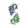
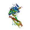
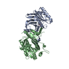
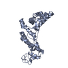
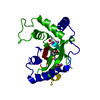
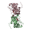
| Title | Structure of the human POT1-TPP1 complex | ||||||
 Components Components |
| ||||||
 Keywords Keywords |  DNA BINDING PROTEIN / POT1 / Telomere DNA BINDING PROTEIN / POT1 / Telomere | ||||||
| Function / homology |  Function and homology information Function and homology informationpositive regulation of single-stranded telomeric DNA binding / positive regulation of DNA strand elongation / regulation of DNA helicase activity / G-rich single-stranded DNA binding / telomere assembly / segmentation / urogenital system development / regulation of double-strand break repair via nonhomologous end joining / protection from non-homologous end joining at telomere / 8-hydroxy-2'-deoxyguanosine DNA binding ...positive regulation of single-stranded telomeric DNA binding / positive regulation of DNA strand elongation / regulation of DNA helicase activity / G-rich single-stranded DNA binding / telomere assembly / segmentation / urogenital system development / regulation of double-strand break repair via nonhomologous end joining / protection from non-homologous end joining at telomere / 8-hydroxy-2'-deoxyguanosine DNA binding / telomeric D-loop binding / positive regulation of helicase activity / telomerase inhibitor activity / DEAD/H-box RNA helicase binding / establishment of protein localization to telomere / positive regulation of DNA helicase activity / telomeric D-loop disassembly / shelterin complex / Telomere C-strand synthesis initiation / regulation of telomere maintenance via telomerase / Telomere C-strand (Lagging Strand) Synthesis / negative regulation of telomerase activity / nuclear telomere cap complex / single-stranded telomeric DNA binding / positive regulation of telomere maintenance / Processive synthesis on the C-strand of the telomere / Polymerase switching on the C-strand of the telomere / Removal of the Flap Intermediate from the C-strand / G-rich strand telomeric DNA binding / telomere capping / telomerase holoenzyme complex / embryonic limb morphogenesis / protein localization to chromosome, telomeric region / DNA duplex unwinding / telomeric DNA binding / negative regulation of telomere maintenance via telomerase / Telomere Extension By Telomerase / telomere maintenance via telomerase / DNA polymerase binding / Packaging Of Telomere Ends / Recognition and association of DNA glycosylase with site containing an affected purine / Cleavage of the damaged purine / positive regulation of telomerase activity / Recognition and association of DNA glycosylase with site containing an affected pyrimidine / Cleavage of the damaged pyrimidine / Inhibition of DNA recombination at telomere / Meiotic synapsis / positive regulation of telomere maintenance via telomerase / telomere maintenance / skeletal system development / intracellular protein transport / DNA Damage/Telomere Stress Induced Senescence / chromosome, telomeric region / nuclear body / protein-containing complex binding / nucleoplasmSimilarity search - Function | ||||||
| Biological species |  Homo sapiens (human) Homo sapiens (human) | ||||||
| Method | X-RAY DIFFRACTION / MOLECULAR REPLACEMENT / Resolution: 2.9 Å | ||||||
 Authors Authors | Aramburu, T. / Skordalakes, E. | ||||||
| Funding support |  United States, 1items United States, 1items
| ||||||
 Citation Citation | Journal: Comput Struct Biotechnol J / Year: 2022 Title: POT1-TPP1 binding stabilizes POT1, promoting efficient telomere maintenance. Authors: Aramburu, T. / Kelich, J. / Rice, C. / Skordalakes, E. | ||||||
| History |
|
- Structure visualization
| Structure viewer | Molecule: MolmilJmol/JSmol |
|---|
- Downloads & links
-Download
| PDBx/mmCIF format | 7s1t.cif.gz | 301.1 KB | Display | PDBx/mmCIF format |
|---|---|---|---|---|
| PDB format | pdb7s1t.ent.gz | 243.1 KB | Display | PDB format |
| PDBx/mmJSON format | 7s1t.json.gz | Tree view | PDBx/mmJSON format | |
| Others |  Other downloads Other downloads |
-Validation report
| Arichive directory | https://data.pdbj.org/pub/pdb/validation_reports/s1/7s1tftp://data.pdbj.org/pub/pdb/validation_reports/s1/7s1t | HTTPS FTP |
|---|
-Related structure data
| Related structure data | 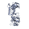 7s1oC  7s1uC 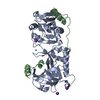 5un7S S: Starting model for refinement C: citing same article ( |
|---|---|
| Similar structure data |

-Links
 PDBj
PDBj


- Assembly
| Deposited unit | 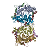
| ||||||||
|---|---|---|---|---|---|---|---|---|---|
| 1 | 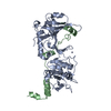
| ||||||||
| 2 | 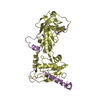
| ||||||||
| 3 | 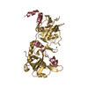
| ||||||||
| 4 | 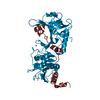
| ||||||||
| Unit cell |
|
-Components
| #1: Protein | Mass: 35446.727 Da / Num. of mol.: 4 Source method: isolated from a genetically manipulated source Source: (gene. exp.) Homo sapiens (human) / Gene: POT1 / Production host:  Escherichia phage EcSzw-2 (virus) / References: UniProt: Q9NUX5 Escherichia phage EcSzw-2 (virus) / References: UniProt: Q9NUX5#2: Protein | Mass: 9841.022 Da / Num. of mol.: 4 Source method: isolated from a genetically manipulated source Source: (gene. exp.) Homo sapiens (human) / Gene: ACD, PIP1, PTOP, TINT1, TPP1 / Production host: Escherichia phage EcSzw-2 (virus) / References: UniProt: Q96AP0#3: Chemical | ChemComp-ZN /   Mass: 65.409 Da / Num. of mol.: 4 / Source method: obtained synthetically / Formula: Zn / Feature type: SUBJECT OF INVESTIGATION Mass: 65.409 Da / Num. of mol.: 4 / Source method: obtained synthetically / Formula: Zn / Feature type: SUBJECT OF INVESTIGATIONHas ligand of interest | Y | |
|---|
-Experimental details
-Experiment
| Experiment | Method: X-RAY DIFFRACTION / Number of used crystals: 1 |
|---|
- Sample preparation
| Crystal | Density Matthews: 2.66 Å3/Da / Density % sol: 53.83 % |
|---|---|
| Crystal grow | Temperature: 290 K / Method: vapor diffusion, sitting drop / Details: 0.1M HEPES pH 7.5 and 3.0M NaCl |
-Data collection
| Diffraction | Mean temperature: 100 K / Serial crystal experiment: N |
|---|---|
| Diffraction source | Source: ROTATING ANODE / Type: RIGAKU MICROMAX-007 / Wavelength: 1.54178 Å |
| Detector | Type: RIGAKU SATURN 944+ / Detector: CCD / Date: Jun 19, 2016 |
| Radiation | Protocol: SINGLE WAVELENGTH / Monochromatic (M) / Laue (L): M / Scattering type: x-ray |
| Radiation wavelength | Wavelength: 1.54178 Å / Relative weight: 1 |
| Reflection | Resolution: 2.9→36.44 Å / Num. obs: 36930 / % possible obs: 90 % / Redundancy: 5.9 % / CC1/2: 0.996 / Net I/σ(I): 9.32 |
| Reflection shell | Resolution: 2.9→2.98 Å / Num. unique obs: 2747 / CC1/2: 0.55 |
- Processing
| Software |
| ||||||||||||||||||||||||||||||||||||||||||||||||||||||||||||||||||||||||||||||||||||||||||||||||||
|---|---|---|---|---|---|---|---|---|---|---|---|---|---|---|---|---|---|---|---|---|---|---|---|---|---|---|---|---|---|---|---|---|---|---|---|---|---|---|---|---|---|---|---|---|---|---|---|---|---|---|---|---|---|---|---|---|---|---|---|---|---|---|---|---|---|---|---|---|---|---|---|---|---|---|---|---|---|---|---|---|---|---|---|---|---|---|---|---|---|---|---|---|---|---|---|---|---|---|---|
| Refinement | Method to determine structure: MOLECULAR REPLACEMENT Starting model: 5UN7 Resolution: 2.9→36.44 Å / SU ML: 0.53 / Cross valid method: THROUGHOUT / σ(F): 1.95 / Phase error: 36.46 / Stereochemistry target values: ML
| ||||||||||||||||||||||||||||||||||||||||||||||||||||||||||||||||||||||||||||||||||||||||||||||||||
| Solvent computation | Shrinkage radii: 0.9 Å / VDW probe radii: 1.11 Å / Solvent model: FLAT BULK SOLVENT MODEL | ||||||||||||||||||||||||||||||||||||||||||||||||||||||||||||||||||||||||||||||||||||||||||||||||||
| Displacement parameters | Biso max: 276.88 Å2 / Biso mean: 76.1356 Å2 / Biso min: 27.36 Å2 | ||||||||||||||||||||||||||||||||||||||||||||||||||||||||||||||||||||||||||||||||||||||||||||||||||
| Refinement step | Cycle: final / Resolution: 2.9→36.44 Å
| ||||||||||||||||||||||||||||||||||||||||||||||||||||||||||||||||||||||||||||||||||||||||||||||||||
| LS refinement shell | Refine-ID: X-RAY DIFFRACTION / Rfactor Rfree error: 0 / Total num. of bins used: 13
|
