 Movie
Movie Controller


+ Open data

- Basic information
| Entry | Database: PDB / ID: 5un7 | ||||||||||||
|---|---|---|---|---|---|---|---|---|---|---|---|---|---|
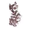
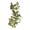
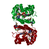
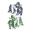
| Title | Structure of the human POT1-TPP1 telomeric complex | ||||||||||||
 Components Components |
| ||||||||||||
 Keywords Keywords |  PROTEIN BINDING / Telomeric DNA binding complex / maintains telomere integrity / DNA BINDING PROTEIN PROTEIN BINDING / Telomeric DNA binding complex / maintains telomere integrity / DNA BINDING PROTEIN | ||||||||||||
| Function / homology |  Function and homology information Function and homology informationpositive regulation of single-stranded telomeric DNA binding / positive regulation of DNA strand elongation / regulation of DNA helicase activity / G-rich single-stranded DNA binding / telomere assembly / segmentation / urogenital system development / regulation of double-strand break repair via nonhomologous end joining / protection from non-homologous end joining at telomere / 8-hydroxy-2'-deoxyguanosine DNA binding ...positive regulation of single-stranded telomeric DNA binding / positive regulation of DNA strand elongation / regulation of DNA helicase activity / G-rich single-stranded DNA binding / telomere assembly / segmentation / urogenital system development / regulation of double-strand break repair via nonhomologous end joining / protection from non-homologous end joining at telomere / 8-hydroxy-2'-deoxyguanosine DNA binding / telomeric D-loop binding / positive regulation of helicase activity / telomerase inhibitor activity / DEAD/H-box RNA helicase binding / establishment of protein localization to telomere / positive regulation of DNA helicase activity / telomeric D-loop disassembly / shelterin complex / Telomere C-strand synthesis initiation / regulation of telomere maintenance via telomerase / Telomere C-strand (Lagging Strand) Synthesis / negative regulation of telomerase activity / nuclear telomere cap complex / single-stranded telomeric DNA binding / positive regulation of telomere maintenance / Processive synthesis on the C-strand of the telomere / Polymerase switching on the C-strand of the telomere / Removal of the Flap Intermediate from the C-strand / G-rich strand telomeric DNA binding / telomere capping / telomerase holoenzyme complex / embryonic limb morphogenesis / protein localization to chromosome, telomeric region / DNA duplex unwinding / telomeric DNA binding / negative regulation of telomere maintenance via telomerase / Telomere Extension By Telomerase / telomere maintenance via telomerase / DNA polymerase binding / Packaging Of Telomere Ends / Recognition and association of DNA glycosylase with site containing an affected purine / Cleavage of the damaged purine / positive regulation of telomerase activity / Recognition and association of DNA glycosylase with site containing an affected pyrimidine / Cleavage of the damaged pyrimidine / Inhibition of DNA recombination at telomere / Meiotic synapsis / positive regulation of telomere maintenance via telomerase / telomere maintenance / skeletal system development / intracellular protein transport / DNA Damage/Telomere Stress Induced Senescence / chromosome, telomeric region / nuclear body / protein-containing complex binding / nucleoplasmSimilarity search - Function | ||||||||||||
| Biological species |  Homo sapiens (human) Homo sapiens (human) | ||||||||||||
| Method | X-RAY DIFFRACTION / SYNCHROTRON / SAD / Resolution: 2.1 Å | ||||||||||||
 Authors Authors | Rice, C. / Doukov, T. / Skordalakes, E. | ||||||||||||
| Funding support |  United States, 3items United States, 3items
| ||||||||||||
 Citation Citation | Journal: Nat Commun / Year: 2017 Title: Structural and functional analysis of the human POT1-TPP1 telomeric complex. Authors: Rice, C. / Shastrula, P.K. / Kossenkov, A.V. / Hills, R. / Baird, D.M. / Showe, L.C. / Doukov, T. / Janicki, S. / Skordalakes, E. | ||||||||||||
| History |
|
- Structure visualization
| Structure viewer | Molecule: MolmilJmol/JSmol |
|---|
- Downloads & links
-Download
| PDBx/mmCIF format | 5un7.cif.gz | 101.1 KB | Display | PDBx/mmCIF format |
|---|---|---|---|---|
| PDB format | pdb5un7.ent.gz | 74.1 KB | Display | PDB format |
| PDBx/mmJSON format | 5un7.json.gz | Tree view | PDBx/mmJSON format | |
| Others |  Other downloads Other downloads |
-Validation report
| Arichive directory | https://data.pdbj.org/pub/pdb/validation_reports/un/5un7ftp://data.pdbj.org/pub/pdb/validation_reports/un/5un7 | HTTPS FTP |
|---|
-Related structure data
| Similar structure data |
|---|


-Links
 PDBj
PDBj


- Assembly
| Deposited unit | 
| ||||||||||||
|---|---|---|---|---|---|---|---|---|---|---|---|---|---|
| 1 |
| ||||||||||||
| 2 | 
| ||||||||||||
| Unit cell |
| ||||||||||||
| Components on special symmetry positions |
|
-Components
| #1: Protein | Mass: 34786.953 Da / Num. of mol.: 1 Source method: isolated from a genetically manipulated source Source: (gene. exp.) Homo sapiens (human) / Gene: POT1 / Production host:  Escherichia coli (E. coli) / References: UniProt: Q9NUX5 Escherichia coli (E. coli) / References: UniProt: Q9NUX5 | ||
|---|---|---|---|
| #2: Protein | Mass: 9195.318 Da / Num. of mol.: 1 / Fragment: UNP residues 255-337 Source method: isolated from a genetically manipulated source Source: (gene. exp.) Homo sapiens (human) / Gene: ACD, PIP1, PTOP, TINT1, TPP1 / Production host: Escherichia coli (E. coli) / References: UniProt: Q96AP0 | ||
| #3: Chemical | ChemComp-ZN /   Mass: 65.409 Da / Num. of mol.: 1 / Source method: obtained synthetically / Formula: Zn Mass: 65.409 Da / Num. of mol.: 1 / Source method: obtained synthetically / Formula: Zn | ||
| #4: Chemical |   Mass: 39.098 Da / Num. of mol.: 3 / Source method: obtained synthetically / Formula: K Mass: 39.098 Da / Num. of mol.: 3 / Source method: obtained synthetically / Formula: K#5: Water | ChemComp-HOH / | Water Mass: 18.015 Da / Num. of mol.: 534 / Source method: isolated from a natural source / Formula: H2O Mass: 18.015 Da / Num. of mol.: 534 / Source method: isolated from a natural source / Formula: H2O |
-Experimental details
-Experiment
| Experiment | Method: X-RAY DIFFRACTION / Number of used crystals: 1 |
|---|
- Sample preparation
| Crystal | Density Matthews: 3.48 Å3/Da / Density % sol: 64.67 % |
|---|---|
| Crystal grow | Temperature: 291 K / Method: vapor diffusion, sitting drop Details: 2.4 M KCl, 50 mM K/Na Tartrate, 20 mM BaCl2, and 0.1 M Sodium Citrate, pH 5.5 |
-Data collection
| Diffraction | Mean temperature: 100 K |
|---|---|
| Diffraction source | Source: SYNCHROTRON / Site: SSRL / Beamline: BL12-2 / Wavelength: 1.03317 Å |
| Detector | Type: DECTRIS PILATUS 6M / Detector: PIXEL / Date: Aug 25, 2016 |
| Radiation | Protocol: SINGLE WAVELENGTH / Monochromatic (M) / Laue (L): M / Scattering type: x-ray |
| Radiation wavelength | Wavelength: 1.03317 Å / Relative weight: 1 |
| Reflection | Resolution: 2.1→38.6 Å / Num. obs: 68409 / % possible obs: 99.8 % / Redundancy: 20 % / CC1/2: 1 / Net I/σ(I): 22.8 |
| Reflection shell | Resolution: 2.1→2.15 Å / Redundancy: 20 % / Mean I/σ(I) obs: 2.7 / Num. unique obs: 5044 / CC1/2: 0.85 / % possible all: 99.8 |
- Processing
| Software |
| ||||||||||||||||||||||||||||||||||||||||||||||||||||||||||||||||||||||||||||||||||||||||||||||||||||||||||||||||||||||||||||||||||||||||||||||||||||||||||||||||||||||||||||||||||||||
|---|---|---|---|---|---|---|---|---|---|---|---|---|---|---|---|---|---|---|---|---|---|---|---|---|---|---|---|---|---|---|---|---|---|---|---|---|---|---|---|---|---|---|---|---|---|---|---|---|---|---|---|---|---|---|---|---|---|---|---|---|---|---|---|---|---|---|---|---|---|---|---|---|---|---|---|---|---|---|---|---|---|---|---|---|---|---|---|---|---|---|---|---|---|---|---|---|---|---|---|---|---|---|---|---|---|---|---|---|---|---|---|---|---|---|---|---|---|---|---|---|---|---|---|---|---|---|---|---|---|---|---|---|---|---|---|---|---|---|---|---|---|---|---|---|---|---|---|---|---|---|---|---|---|---|---|---|---|---|---|---|---|---|---|---|---|---|---|---|---|---|---|---|---|---|---|---|---|---|---|---|---|---|---|
| Refinement | Method to determine structure: SAD / Resolution: 2.1→20 Å / Cor.coef. Fo:Fc: 0.962 / Cor.coef. Fo:Fc free: 0.945 / SU B: 3.842 / SU ML: 0.101 / Cross valid method: THROUGHOUT / ESU R: 0.156 / ESU R Free: 0.148 / Stereochemistry target values: MAXIMUM LIKELIHOOD
| ||||||||||||||||||||||||||||||||||||||||||||||||||||||||||||||||||||||||||||||||||||||||||||||||||||||||||||||||||||||||||||||||||||||||||||||||||||||||||||||||||||||||||||||||||||||
| Solvent computation | Ion probe radii: 0.8 Å / Shrinkage radii: 0.8 Å / VDW probe radii: 1.2 Å / Solvent model: BABINET MODEL WITH MASK | ||||||||||||||||||||||||||||||||||||||||||||||||||||||||||||||||||||||||||||||||||||||||||||||||||||||||||||||||||||||||||||||||||||||||||||||||||||||||||||||||||||||||||||||||||||||
| Displacement parameters | Biso mean: 42.064 Å2
| ||||||||||||||||||||||||||||||||||||||||||||||||||||||||||||||||||||||||||||||||||||||||||||||||||||||||||||||||||||||||||||||||||||||||||||||||||||||||||||||||||||||||||||||||||||||
| Refinement step | Cycle: 1 / Resolution: 2.1→20 Å
| ||||||||||||||||||||||||||||||||||||||||||||||||||||||||||||||||||||||||||||||||||||||||||||||||||||||||||||||||||||||||||||||||||||||||||||||||||||||||||||||||||||||||||||||||||||||
| Refine LS restraints |
|