 Movie
Movie Controller


+ Open data

- Basic information
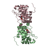
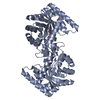
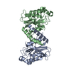
| Entry | Database: PDB / ID: 1jq6 | ||||||
|---|---|---|---|---|---|---|---|
| Title | HUMAN CYTOMEGALOVIRUS PROTEASE DIMER-INTERFACE MUTANT, S225Y | ||||||
 Components Components | ASSEMBLIN | ||||||
 Keywords Keywords | HYDROLASE / Herpesvirus / cytomegalovirus / serine protease / dimerization / enzyme activity regulation | ||||||
| Function / homology |  Function and homology informationassemblin / viral release from host cell / host cell cytoplasm / serine-type endopeptidase activity / host cell nucleus / proteolysis / identical protein binding Function and homology informationassemblin / viral release from host cell / host cell cytoplasm / serine-type endopeptidase activity / host cell nucleus / proteolysis / identical protein bindingSimilarity search - Function | ||||||
| Biological species |   Human herpesvirus 5 Human herpesvirus 5 | ||||||
| Method | X-RAY DIFFRACTION / SYNCHROTRON / MOLECULAR REPLACEMENT / Resolution: 2.3 Å | ||||||
 Authors Authors | Batra, R. / Khayat, R. / Tong, L. | ||||||
 Citation Citation | Journal: Nat.Struct.Biol. / Year: 2001 Title: Molecular mechanism for dimerization to regulate the catalytic activity of human cytomegalovirus protease. Authors: Batra, R. / Khayat, R. / Tong, L. | ||||||
| History |
|
- Structure visualization
| Structure viewer | Molecule: MolmilJmol/JSmol |
|---|
- Downloads & links
-Download
| PDBx/mmCIF format | 1jq6.cif.gz | 49.2 KB | Display | PDBx/mmCIF format |
|---|---|---|---|---|
| PDB format | pdb1jq6.ent.gz | 33.6 KB | Display | PDB format |
| PDBx/mmJSON format | 1jq6.json.gz | Tree view | PDBx/mmJSON format | |
| Others |  Other downloads Other downloads |
-Validation report
| Arichive directory | https://data.pdbj.org/pub/pdb/validation_reports/jq/1jq6ftp://data.pdbj.org/pub/pdb/validation_reports/jq/1jq6 | HTTPS FTP |
|---|
-Related structure data
| Related structure data |  1jq7C  1wpoS S: Starting model for refinement C: citing same article ( |
|---|---|
| Similar structure data |


-Links
 PDBj
PDBj- Assembly
| Deposited unit | 
| ||||||||
|---|---|---|---|---|---|---|---|---|---|
| 1 | 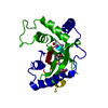
| ||||||||
| Unit cell |
| ||||||||
| Details | Monomer in the asymmetric unit, but biological assembly unit is a dimer that can be generated by the two fold axis. |
-Components
| #1: Protein | / PROTEASE Mass: 28518.590 Da / Num. of mol.: 1 / Mutation: A143Q, S225Y Source method: isolated from a genetically manipulated source Source: (gene. exp.) Human herpesvirus 5 / Genus: Cytomegalovirus / Production host:  Escherichia coli (E. coli) / References: UniProt: P16753, assemblin Escherichia coli (E. coli) / References: UniProt: P16753, assemblin |
|---|---|
| #2: Water | ChemComp-HOH / Water Mass: 18.015 Da / Num. of mol.: 42 / Source method: isolated from a natural source / Formula: H2O Mass: 18.015 Da / Num. of mol.: 42 / Source method: isolated from a natural source / Formula: H2O |
-Experimental details
-Experiment
| Experiment | Method: X-RAY DIFFRACTION / Number of used crystals: 1 |
|---|
- Sample preparation
| Crystal | Density Matthews: 2.18 Å3/Da / Density % sol: 43.47 % | ||||||||||||||||||||||||||||||||||||||||||||||||||||||||||||||||||
|---|---|---|---|---|---|---|---|---|---|---|---|---|---|---|---|---|---|---|---|---|---|---|---|---|---|---|---|---|---|---|---|---|---|---|---|---|---|---|---|---|---|---|---|---|---|---|---|---|---|---|---|---|---|---|---|---|---|---|---|---|---|---|---|---|---|---|---|
| Crystal grow | Temperature: 294 K / Method: vapor diffusion, sitting drop / pH: 7.2 Details: PEG 8000, sodium cacodylate, magnesium acetate, glycerol, spermine tetrahydrochloride, DTT, pH 7.2, VAPOR DIFFUSION, SITTING DROP, temperature 294K | ||||||||||||||||||||||||||||||||||||||||||||||||||||||||||||||||||
| Crystal grow | *PLUS Temperature: 21 ℃ / pH: 5 | ||||||||||||||||||||||||||||||||||||||||||||||||||||||||||||||||||
| Components of the solutions | *PLUS
|
-Data collection
| Diffraction | Mean temperature: 100 K |
|---|---|
| Diffraction source | Source: SYNCHROTRON / Site: NSLS  / Beamline: X4A / Wavelength: 0.976 Å / Beamline: X4A / Wavelength: 0.976 Å |
| Detector | Type: FUJI / Detector: IMAGE PLATE / Date: Nov 9, 2000 |
| Radiation | Monochromator: Si 111 Channel / Protocol: SINGLE WAVELENGTH / Monochromatic (M) / Laue (L): M / Scattering type: x-ray |
| Radiation wavelength | Wavelength: 0.976 Å / Relative weight: 1 |
| Reflection | Resolution: 2.3→19.3 Å / Num. all: 38153 / Num. obs: 10420 / % possible obs: 91 % / Observed criterion σ(F): 2 / Observed criterion σ(I): 2 / Biso Wilson estimate: 18.5 Å2 |
| Reflection shell | Resolution: 2.3→2.44 Å / % possible all: 93 |
| Reflection | *PLUS % possible obs: 91 % / Num. measured all: 38153 / Rmerge(I) obs: 0.057 |
| Reflection shell | *PLUS Rmerge(I) obs: 0.172 |
- Processing
| Software |
| ||||||||||||||||||||||||||||||||||||||||
|---|---|---|---|---|---|---|---|---|---|---|---|---|---|---|---|---|---|---|---|---|---|---|---|---|---|---|---|---|---|---|---|---|---|---|---|---|---|---|---|---|---|
| Refinement | Method to determine structure: MOLECULAR REPLACEMENT Starting model: PDB ENTRY 1WPO (monomer) Resolution: 2.3→19.3 Å / Rfactor Rfree error: 0.01 / Data cutoff high absF: 5293688.41 / Data cutoff low absF: 0 / Isotropic thermal model: RESTRAINED / Cross valid method: THROUGHOUT / σ(F): 2 / Stereochemistry target values: Engh & Huber
| ||||||||||||||||||||||||||||||||||||||||
| Solvent computation | Solvent model: FLAT MODEL / Bsol: 56.4329 Å2 / ksol: 0.394661 e/Å3 | ||||||||||||||||||||||||||||||||||||||||
| Displacement parameters | Biso mean: 43.2 Å2
| ||||||||||||||||||||||||||||||||||||||||
| Refine analyze |
| ||||||||||||||||||||||||||||||||||||||||
| Refinement step | Cycle: LAST / Resolution: 2.3→19.3 Å
| ||||||||||||||||||||||||||||||||||||||||
| Refine LS restraints |
| ||||||||||||||||||||||||||||||||||||||||
| LS refinement shell | Resolution: 2.3→2.44 Å / Rfactor Rfree error: 0.026 / Total num. of bins used: 6
| ||||||||||||||||||||||||||||||||||||||||
| Xplor file |
| ||||||||||||||||||||||||||||||||||||||||
| Software | *PLUS Name: CNS / Version: 1 / Classification: refinement | ||||||||||||||||||||||||||||||||||||||||
| Refinement | *PLUS σ(F): 2 / % reflection Rfree: 7.6 % / Rfactor obs: 0.229 | ||||||||||||||||||||||||||||||||||||||||
| Solvent computation | *PLUS | ||||||||||||||||||||||||||||||||||||||||
| Displacement parameters | *PLUS Biso mean: 43.2 Å2 | ||||||||||||||||||||||||||||||||||||||||
| Refine LS restraints | *PLUS
| ||||||||||||||||||||||||||||||||||||||||
| LS refinement shell | *PLUS Rfactor Rfree: 0.284 / % reflection Rfree: 7.5 % / Rfactor Rwork: 0.249 |
