 Movie
Movie Controller


+ Open data

- Basic information
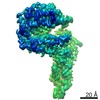
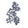
| Entry | Database: PDB / ID: 7qla | |||||||||
|---|---|---|---|---|---|---|---|---|---|---|
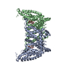
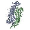
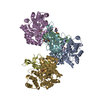
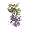
| Title | Structure of the Rab GEF complex Mon1-Ccz1 | |||||||||
 Components Components |
| |||||||||
 Keywords Keywords |  ENDOCYTOSIS / guanine nucleotide exchange factor / TLD Rab GEF / longin domains / PIP binding ENDOCYTOSIS / guanine nucleotide exchange factor / TLD Rab GEF / longin domains / PIP binding | |||||||||
| Function / homology |  Function and homology information Function and homology informationprotein targeting to vacuole / multivesicular body membrane / vacuolar membrane / vesicle-mediated transport / autophagySimilarity search - Function | |||||||||
| Biological species |  Chaetomium thermophilum (fungus) Chaetomium thermophilum (fungus) | |||||||||
| Method | ELECTRON MICROSCOPY / single particle reconstruction / cryo EM / Resolution: 3.85 Å | |||||||||
 Authors Authors | Klink, B.U. / Herrmann, E. / Antoni, C. / Langemeyer, L. / Kiontke, S. / Gatsogiannis, C. / Ungermann, C. / Raunser, S. / Kuemmel, D. | |||||||||
| Funding support |  Germany, 2items Germany, 2items
| |||||||||
 Citation Citation | Journal: Proc Natl Acad Sci U S A / Year: 2022 Title: Structure of the Mon1-Ccz1 complex reveals molecular basis of membrane binding for Rab7 activation. Authors: Björn U Klink / Eric Herrmann / Claudia Antoni / Lars Langemeyer / Stephan Kiontke / Christos Gatsogiannis / Christian Ungermann / Stefan Raunser / Daniel Kümmel / Abstract: Activation of the GTPase Rab7/Ypt7 by its cognate guanine nucleotide exchange factor (GEF) Mon1-Ccz1 marks organelles such as endosomes and autophagosomes for fusion with lysosomes/vacuoles and ...Activation of the GTPase Rab7/Ypt7 by its cognate guanine nucleotide exchange factor (GEF) Mon1-Ccz1 marks organelles such as endosomes and autophagosomes for fusion with lysosomes/vacuoles and degradation of their content. Here, we present a high-resolution cryogenic electron microscopy structure of the Mon1-Ccz1 complex that reveals its architecture in atomic detail. Mon1 and Ccz1 are arranged side by side in a pseudo-twofold symmetrical heterodimer. The three Longin domains of each Mon1 and Ccz1 are triangularly arranged, providing a strong scaffold for the catalytic center of the GEF. At the opposite side of the Ypt7-binding site, a positively charged and relatively flat patch stretches the Longin domains 2/3 of Mon1 and functions as a phosphatidylinositol phosphate-binding site, explaining how the GEF is targeted to membranes. Our work provides molecular insight into the mechanisms of endosomal Rab activation and serves as a blueprint for understanding the function of members of the Tri Longin domain Rab-GEF family. | |||||||||
| History |
|
- Structure visualization
| Movie |
Movie viewer |
|---|---|
| Structure viewer | Molecule: MolmilJmol/JSmol |
- Downloads & links
-Download
| PDBx/mmCIF format | 7qla.cif.gz | 164.8 KB | Display | PDBx/mmCIF format |
|---|---|---|---|---|
| PDB format | pdb7qla.ent.gz | 129.2 KB | Display | PDB format |
| PDBx/mmJSON format | 7qla.json.gz | Tree view | PDBx/mmJSON format | |
| Others |  Other downloads Other downloads |
-Validation report
| Arichive directory | https://data.pdbj.org/pub/pdb/validation_reports/ql/7qlaftp://data.pdbj.org/pub/pdb/validation_reports/ql/7qla | HTTPS FTP |
|---|
-Related structure data
| Related structure data | 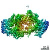 14066MC M: map data used to model this data C: citing same article ( |
|---|---|
| Similar structure data |

-Links
 PDBj
PDBj- Assembly
| Deposited unit | 
|
|---|---|
| 1 |
|
-Components
| #1: Protein | Mass: 57571.832 Da / Num. of mol.: 1 Source method: isolated from a genetically manipulated source Source: (gene. exp.) Chaetomium thermophilum (fungus) / Strain: DSM 1495 / CBS 144.50 / IMI 039719 / Gene: CTHT_0067370 / Production host:  Escherichia coli (E. coli) / References: UniProt: G0SGS3 Escherichia coli (E. coli) / References: UniProt: G0SGS3 |
|---|---|
| #2: Protein | Mass: 75780.461 Da / Num. of mol.: 1 Source method: isolated from a genetically manipulated source Source: (gene. exp.) Chaetomium thermophilum (fungus) / Strain: DSM 1495 / CBS 144.50 / IMI 039719 / Gene: CTHT_0067370 / Production host: Escherichia coli (E. coli) |
-Experimental details
-Experiment
| Experiment | Method: ELECTRON MICROSCOPY |
|---|---|
| EM experiment | Aggregation state: PARTICLE / 3D reconstruction method: single particle reconstruction |
- Sample preparation
| Component | Name: Rab GEF complex Mon1-Ccz1 / Type: COMPLEX / Details: CtMon1 aa141-665; CtCcz1 aa1-796delta361-460 / Entity ID: all / Source: RECOMBINANT | |||||||||||||||||||||||||
|---|---|---|---|---|---|---|---|---|---|---|---|---|---|---|---|---|---|---|---|---|---|---|---|---|---|---|
| Molecular weight | Experimental value: NO | |||||||||||||||||||||||||
| Source (natural) | Organism: Chaetomium thermophilum (fungus) | |||||||||||||||||||||||||
| Source (recombinant) | Organism: Escherichia coli (E. coli) / Strain: BL21 | |||||||||||||||||||||||||
| Buffer solution | pH: 7.3 | |||||||||||||||||||||||||
| Buffer component |
| |||||||||||||||||||||||||
| Specimen | Embedding applied: NO / Shadowing applied: NO / Staining applied: NO / Vitrification applied: YES | |||||||||||||||||||||||||
| Specimen support | Grid material: COPPER / Grid type: Quantifoil R2/1 | |||||||||||||||||||||||||
| Vitrification | Instrument: FEI VITROBOT MARK II / Cryogen name: ETHANE / Humidity: 100 % |
- Electron microscopy imaging
| Experimental equipment |  Model: Titan Krios / Image courtesy: FEI Company |
|---|---|
| Microscopy | Model: FEI TITAN KRIOS |
| Electron gun | Electron source: FIELD EMISSION GUN / Accelerating voltage: 300 kV / Illumination mode: SPOT SCAN |
| Electron lens | Mode: BRIGHT FIELDBright-field microscopy / Cs: 0.001 mm |
| Specimen holder | Cryogen: NITROGEN / Specimen holder model: FEI TITAN KRIOS AUTOGRID HOLDER |
| Image recording | Average exposure time: 15 sec. / Electron dose: 73 e/Å2 / Detector mode: COUNTING / Film or detector model: GATAN K2 SUMMIT (4k x 4k) / Num. of grids imaged: 1 / Num. of real images: 11916 Details: Images were collected in movie-mode with 4 frames per second |
| EM imaging optics | Energyfilter slit width: 20 eV / Phase plate: VOLTA PHASE PLATE |
| Image scans | Movie frames/image: 60 |
- Processing
| Software | Name: PHENIX / Version: 1.19.1_4122: / Classification: refinement | ||||||||||||||||||||||||
|---|---|---|---|---|---|---|---|---|---|---|---|---|---|---|---|---|---|---|---|---|---|---|---|---|---|
| EM software |
| ||||||||||||||||||||||||
| CTF correction | Type: PHASE FLIPPING AND AMPLITUDE CORRECTION | ||||||||||||||||||||||||
| Particle selection | Num. of particles selected: 7155250 | ||||||||||||||||||||||||
| 3D reconstruction | Resolution: 3.85 Å / Resolution method: FSC 0.143 CUT-OFF / Num. of particles: 911674 / Num. of class averages: 1 / Symmetry type: POINT | ||||||||||||||||||||||||
| Atomic model building | Protocol: AB INITIO MODEL | ||||||||||||||||||||||||
| Refine LS restraints |
|
