 Movie
Movie Controller

[English] 日本語
 Yorodumi
- PDB-7ov2: Crystal structure of pig purple acid phosphatase in complex with ... -
+ Open data

- Basic information
| Entry | Database: PDB / ID: 7ov2 | |||||||||
|---|---|---|---|---|---|---|---|---|---|---|
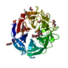
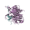
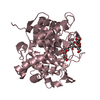
| Title | Crystal structure of pig purple acid phosphatase in complex with L-glutamine, (poly)ethylene glycol fragments and glycerol | |||||||||
 Components Components | Tartrate-resistant acid phosphatase type 5 | |||||||||
 Keywords Keywords |  HYDROLASE / Purple acid phosphatase / metallohydrolases / osteoporosis / fragment-based drug design / METAL BINDING PROTEIN HYDROLASE / Purple acid phosphatase / metallohydrolases / osteoporosis / fragment-based drug design / METAL BINDING PROTEIN | |||||||||
| Function / homology |  Function and homology information Function and homology informationVitamin B2 (riboflavin) metabolism / acid phosphatase / acid phosphatase activity / bone resorption / ferric iron binding / ferrous iron binding / iron ion transport / extracellular regionSimilarity search - Function | |||||||||
| Biological species |  Sus scrofa (pig) Sus scrofa (pig) | |||||||||
| Method | X-RAY DIFFRACTION / SYNCHROTRON / MOLECULAR REPLACEMENT / Resolution: 2.1 Å | |||||||||
 Authors Authors | Feder, D. / McGeary, R.P. / Guddat, L.W. / Schenk, G. | |||||||||
| Funding support |  Australia, 2items Australia, 2items
| |||||||||
 Citation Citation | Journal: Chemmedchem / Year: 2021 Title: Rational Design of Potent Inhibitors of a Metallohydrolase Using a Fragment-Based Approach. Authors: Feder, D. / Mohd-Pahmi, S.H. / Hussein, W.M. / Guddat, L.W. / McGeary, R.P. / Schenk, G. | |||||||||
| History |
|
- Structure visualization
| Structure viewer | Molecule: MolmilJmol/JSmol |
|---|
- Downloads & links
-Download
| PDBx/mmCIF format | 7ov2.cif.gz | 125.4 KB | Display | PDBx/mmCIF format |
|---|---|---|---|---|
| PDB format | pdb7ov2.ent.gz | 78.1 KB | Display | PDB format |
| PDBx/mmJSON format | 7ov2.json.gz | Tree view | PDBx/mmJSON format | |
| Others |  Other downloads Other downloads |
-Validation report
| Arichive directory | https://data.pdbj.org/pub/pdb/validation_reports/ov/7ov2ftp://data.pdbj.org/pub/pdb/validation_reports/ov/7ov2 | HTTPS FTP |
|---|
-Related structure data
| Related structure data |  7ov3C 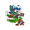 7ov8C  1uteS  6vkb S: Starting model for refinement C: citing same article ( |
|---|---|
| Similar structure data |


-Links
 PDBj
PDBj
- Assembly
| Deposited unit | 
| ||||||||||||
|---|---|---|---|---|---|---|---|---|---|---|---|---|---|
| 1 |
| ||||||||||||
| Unit cell |
|
-Components
-Protein / Sugars , 2 types, 2 molecules A
| #1: Protein | Mass: 35154.066 Da / Num. of mol.: 1 / Source method: isolated from a natural source / Source: (natural) Sus scrofa (pig) / References: UniProt: P09889, acid phosphatase |
|---|---|
| #2: Polysaccharide | beta-D-mannopyranose-(1-3)-beta-D-mannopyranose-(1-4)-2-acetamido-2-deoxy-beta-D-glucopyranose-(1-4) ...beta-D-mannopyranose-(1-3)-beta-D-mannopyranose-(1-4)-2-acetamido-2-deoxy-beta-D-glucopyranose-(1-4)-2-acetamido-2-deoxy-beta-D-glucopyranose / Mass: 748.682 Da / Num. of mol.: 1 Source method: isolated from a genetically manipulated source |
-Non-polymers , 12 types, 276 molecules 


| #3: Chemical | Polyethylene glycol Mass: 238.278 Da / Num. of mol.: 2 / Source method: obtained synthetically / Formula: C10H22O6 / Comment: precipitant*YM Mass: 238.278 Da / Num. of mol.: 2 / Source method: obtained synthetically / Formula: C10H22O6 / Comment: precipitant*YM#4: Chemical | ChemComp-P6G / | Polyethylene glycol Mass: 282.331 Da / Num. of mol.: 1 / Source method: obtained synthetically / Formula: C12H26O7 / Comment: precipitant*YM Mass: 282.331 Da / Num. of mol.: 1 / Source method: obtained synthetically / Formula: C12H26O7 / Comment: precipitant*YM#5: Chemical | Polyethylene glycol Mass: 194.226 Da / Num. of mol.: 2 / Source method: obtained synthetically / Formula: C8H18O5 / Comment: precipitant*YM Mass: 194.226 Da / Num. of mol.: 2 / Source method: obtained synthetically / Formula: C8H18O5 / Comment: precipitant*YM#6: Chemical | ChemComp-GLN / Glutamine Type: L-peptide linking / Mass: 146.144 Da / Num. of mol.: 28 / Source method: obtained synthetically / Formula: C5H10N2O3 / Feature type: SUBJECT OF INVESTIGATION Type: L-peptide linking / Mass: 146.144 Da / Num. of mol.: 28 / Source method: obtained synthetically / Formula: C5H10N2O3 / Feature type: SUBJECT OF INVESTIGATION#7: Chemical | ChemComp-PO4 / | Phosphate Mass: 94.971 Da / Num. of mol.: 1 Mass: 94.971 Da / Num. of mol.: 1Source method: isolated from a genetically manipulated source Formula: PO4 #8: Chemical | ChemComp-EDO / Ethylene glycol Mass: 62.068 Da / Num. of mol.: 17 / Source method: obtained synthetically / Formula: C2H6O2 Mass: 62.068 Da / Num. of mol.: 17 / Source method: obtained synthetically / Formula: C2H6O2#9: Chemical | ChemComp-PGE / Polyethylene glycol Mass: 150.173 Da / Num. of mol.: 8 / Source method: obtained synthetically / Formula: C6H14O4 Mass: 150.173 Da / Num. of mol.: 8 / Source method: obtained synthetically / Formula: C6H14O4#10: Chemical | ChemComp-GOL / Glycerol Mass: 92.094 Da / Num. of mol.: 13 / Source method: obtained synthetically / Formula: C3H8O3 Mass: 92.094 Da / Num. of mol.: 13 / Source method: obtained synthetically / Formula: C3H8O3#11: Chemical | ChemComp-CIT / | Citric acid Mass: 192.124 Da / Num. of mol.: 1 / Source method: obtained synthetically / Formula: C6H8O7 Mass: 192.124 Da / Num. of mol.: 1 / Source method: obtained synthetically / Formula: C6H8O7#12: Chemical | ChemComp-NA / |  Mass: 22.990 Da / Num. of mol.: 1 / Source method: obtained synthetically / Formula: Na Mass: 22.990 Da / Num. of mol.: 1 / Source method: obtained synthetically / Formula: Na#13: Chemical | Iron Mass: 55.845 Da / Num. of mol.: 2 / Source method: obtained synthetically / Formula: Fe Mass: 55.845 Da / Num. of mol.: 2 / Source method: obtained synthetically / Formula: Fe#14: Water | ChemComp-HOH / | WaterMass: 18.015 Da / Num. of mol.: 200 / Source method: isolated from a natural source / Formula: H2O |
|---|
-Details
| Has ligand of interest | Y |
|---|
-Experimental details
-Experiment
| Experiment | Method: X-RAY DIFFRACTION / Number of used crystals: 1 |
|---|
- Sample preparation
| Crystal | Density Matthews: 2.44 Å3/Da / Density % sol: 49.51 % / Description: Rod-shaped purple crystals |
|---|---|
| Crystal grow | Temperature: 291.15 K / Method: vapor diffusion, sitting drop / pH: 5 Details: 25% (W/V) PEG3350, 0.1 M LICL, 5% (V/V)ISOPROPANOL, 0.1 M SODIUM CITRATE PH 5.0, PROTEIN CONCENTRATION 38 MG/ML |
-Data collection
| Diffraction | Mean temperature: 100 K / Serial crystal experiment: N |
|---|---|
| Diffraction source | Source: SYNCHROTRON / Site: Australian Synchrotron / Beamline: MX2 / Wavelength: 1 Å |
| Detector | Type: ADSC QUANTUM 315r / Detector: CCD / Date: Nov 11, 2012 |
| Radiation | Protocol: SINGLE WAVELENGTH / Monochromatic (M) / Laue (L): M / Scattering type: x-ray |
| Radiation wavelength | Wavelength: 1 Å / Relative weight: 1 |
| Reflection | Resolution: 2.1→77.01 Å / Num. obs: 20575 / % possible obs: 100 % / Redundancy: 7.2 % / Biso Wilson estimate: 19.98 Å2 / CC1/2: 0.997 / Rmerge(I) obs: 0.131 / Rpim(I) all: 0.052 / Rrim(I) all: 0.141 / Net I/σ(I): 12.8 |
| Reflection shell | Resolution: 2.1→2.21 Å / Rmerge(I) obs: 0.491 / Mean I/σ(I) obs: 4.4 / Num. unique obs: 2938 / CC1/2: 0.905 / Rpim(I) all: 0.193 / Rrim(I) all: 0.528 / % possible all: 100 |
- Processing
| Software |
| ||||||||||||||||||||||||||||||||||||||||||||||||||||||||
|---|---|---|---|---|---|---|---|---|---|---|---|---|---|---|---|---|---|---|---|---|---|---|---|---|---|---|---|---|---|---|---|---|---|---|---|---|---|---|---|---|---|---|---|---|---|---|---|---|---|---|---|---|---|---|---|---|---|
| Refinement | Method to determine structure: MOLECULAR REPLACEMENT Starting model: 1UTE Resolution: 2.1→51.25 Å / SU ML: 0.1711 / Cross valid method: FREE R-VALUE / σ(F): 1.37 / Phase error: 16.7341
| ||||||||||||||||||||||||||||||||||||||||||||||||||||||||
| Solvent computation | Shrinkage radii: 0.9 Å / VDW probe radii: 1.11 Å | ||||||||||||||||||||||||||||||||||||||||||||||||||||||||
| Displacement parameters | Biso mean: 27.55 Å2 | ||||||||||||||||||||||||||||||||||||||||||||||||||||||||
| Refinement step | Cycle: LAST / Resolution: 2.1→51.25 Å
| ||||||||||||||||||||||||||||||||||||||||||||||||||||||||
| Refine LS restraints |
| ||||||||||||||||||||||||||||||||||||||||||||||||||||||||
| LS refinement shell |
|
