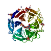
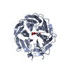
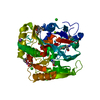
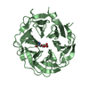
Entry Database : PDB / ID : 2w5oTitle Complex structure of the GH93 alpha-L-arabinofuranosidase of Fusarium graminearum with arabinobiose ALPHA-L-ARABINOFURANOSIDASE Keywords / / / / Function / homology / / / / Biological species GIBBERELLA ZEAE (fungus)Method / / / Resolution : 2.05 Å Authors Carapito, R. / Imberty, A. / Jeltsch, J.M. / Byrns, S.C. / Tam, P.H. / Lowary, T.L. / Varrot, A. / Phalip, V. Journal : J.Biol.Chem. / Year : 2009Title : Molecular Basis of Arabinobio-Hydrolase Activity in Phytopathogenic Fungi. Crystal Structure and Catalytic Mechanism of Fusarium Graminearum Gh93 Exo-Alpha-L-Arabinanase.Authors : Carapito, R. / Imberty, A. / Jeltsch, J.M. / Byrns, S.C. / Tam, P.H. / Lowary, T.L. / Varrot, A. / Phalip, V. History Deposition Dec 11, 2008 Deposition site / Processing site Revision 1.0 Mar 3, 2009 Provider / Type Revision 1.1 May 8, 2011 Group Revision 1.2 Jul 13, 2011 Group Revision 2.0 Jul 29, 2020 Group Atomic model / Data collection ... Atomic model / Data collection / Derived calculations / Other / Structure summary Category atom_site / chem_comp ... atom_site / chem_comp / entity / pdbx_branch_scheme / pdbx_chem_comp_identifier / pdbx_database_status / pdbx_entity_branch / pdbx_entity_branch_descriptor / pdbx_entity_branch_link / pdbx_entity_branch_list / pdbx_entity_nonpoly / pdbx_nonpoly_scheme / pdbx_struct_assembly_gen / struct_asym / struct_conn / struct_site / struct_site_gen Item _atom_site.auth_asym_id / _atom_site.auth_atom_id ... _atom_site.auth_asym_id / _atom_site.auth_atom_id / _atom_site.auth_seq_id / _atom_site.label_asym_id / _atom_site.label_atom_id / _chem_comp.name / _chem_comp.type / _entity.formula_weight / _entity.pdbx_description / _entity.pdbx_number_of_molecules / _entity.type / _pdbx_database_status.status_code_sf / _pdbx_struct_assembly_gen.asym_id_list / _struct_conn.pdbx_leaving_atom_flag / _struct_conn.ptnr1_auth_asym_id / _struct_conn.ptnr1_auth_seq_id / _struct_conn.ptnr1_label_atom_id / _struct_conn.ptnr2_auth_asym_id / _struct_conn.ptnr2_auth_seq_id / _struct_conn.ptnr2_label_asym_id / _struct_conn.ptnr2_label_atom_id Description / Provider / Type Revision 2.1 Dec 13, 2023 Group Data collection / Database references ... Data collection / Database references / Derived calculations / Refinement description / Structure summary Category chem_comp / chem_comp_atom ... chem_comp / chem_comp_atom / chem_comp_bond / database_2 / pdbx_initial_refinement_model / struct_conn Item _chem_comp.pdbx_synonyms / _database_2.pdbx_DOI ... _chem_comp.pdbx_synonyms / _database_2.pdbx_DOI / _database_2.pdbx_database_accession / _struct_conn.pdbx_leaving_atom_flag
Show all Show less Remark 650 HELIX DETERMINATION METHOD: AUTHOR PROVIDED. Remark 700 SHEET DETERMINATION METHOD: AUTHOR PROVIDED.
 Movie
Movie Controller

 Yorodumi
 Open data
 Basic information
 Components
 Keywords
 Function and homology information
 Authors
 Citation
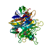
 Structure visualization
 Downloads & links
 Other downloads
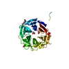


 PDBj
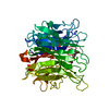
PDBj Assembly
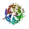


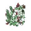
 Mass: 62.068 Da / Num. of mol.: 5 / Source method: obtained synthetically / Formula: C2H6O2
 Mass: 96.063 Da / Num. of mol.: 1 / Source method: obtained synthetically / Formula: SO4
 Mass: 106.120 Da / Num. of mol.: 1 / Source method: obtained synthetically / Formula: C4H10O3
 Sample preparation
 / Beamline: ID14-2 / Wavelength: 0.933
  Processing