 Movie
Movie Controller


+ Open data

- Basic information
| Entry | Database: PDB / ID: 1qjw | |||||||||
|---|---|---|---|---|---|---|---|---|---|---|
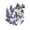
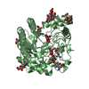
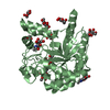
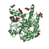
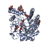
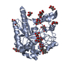
| Title | CEL6A (Y169F) WITH A NON-HYDROLYSABLE CELLOTETRAOSE | |||||||||
 Components Components | CELLOBIOHYDROLASE CEL6A (FORMERLY CALLED CBH II) | |||||||||
 Keywords Keywords |  HYDROLASE / GLYCOSIDASE / GLYCOPROTEIN HYDROLASE / GLYCOSIDASE / GLYCOPROTEIN | |||||||||
| Function / homology |  Function and homology information Function and homology informationcellulose 1,4-beta-cellobiosidase (non-reducing end) / cellulose 1,4-beta-cellobiosidase activity / cellulose binding / cellulose catabolic process / extracellular region / identical protein bindingSimilarity search - Function | |||||||||
| Biological species |  TRICHODERMA REESEI (fungus) TRICHODERMA REESEI (fungus) | |||||||||
| Method | X-RAY DIFFRACTION / MOLECULAR REPLACEMENT / Resolution: 1.9 Å | |||||||||
 Authors Authors | Zou, J.-Y. / Jones, T.A. | |||||||||
 Citation Citation | Journal: Structure / Year: 1999 Title: Crystallographic Evidence for Substrate Ring Distortion and Protein Conformational Changes During Catalysis in Cellobiohydrolase Cel6A from Trichoderma Reesei Authors: Zou, J.-Y. / Kleywegt, G.J. / Stahlberg, J. / Driguez, H. / Nerinckx, W. / Claeyssens, M. / Koivula, A. / Teeri, T.T. / Jones, T.A. #1: Journal: Science / Year: 1990Title: Three-Dimensional Structure of Cellobiohydrolase II from Trichoderma Reesei Authors: Rouvinen, J. / Bergfors, T. / Teeri, T. / Knowles, J.K. / Jones, T.A. | |||||||||
| History |
|
- Structure visualization
| Structure viewer | Molecule: MolmilJmol/JSmol |
|---|
- Downloads & links
-Download
| PDBx/mmCIF format | 1qjw.cif.gz | 167.2 KB | Display | PDBx/mmCIF format |
|---|---|---|---|---|
| PDB format | pdb1qjw.ent.gz | 134.9 KB | Display | PDB format |
| PDBx/mmJSON format | 1qjw.json.gz | Tree view | PDBx/mmJSON format | |
| Others |  Other downloads Other downloads |
-Validation report
| Arichive directory | https://data.pdbj.org/pub/pdb/validation_reports/qj/1qjwftp://data.pdbj.org/pub/pdb/validation_reports/qj/1qjw | HTTPS FTP |
|---|
-Related structure data


-Links
 PDBj
PDBj


- Assembly
| Deposited unit | 
| ||||||||
|---|---|---|---|---|---|---|---|---|---|
| 1 | 
| ||||||||
| 2 | 
| ||||||||
| Unit cell |
| ||||||||
| Noncrystallographic symmetry (NCS) | NCS oper: (Code: given Matrix: (0.999999, -0.001278, -0.000251), Vector : |
-Components
-Protein , 1 types, 2 molecules AB
| #1: Protein | Mass: 39039.297 Da / Num. of mol.: 2 / Fragment: CATALYTIC DOMAIN, RESIDUES 83-447 / Mutation: YES Source method: isolated from a genetically manipulated source Source: (gene. exp.) TRICHODERMA REESEI (fungus) / Gene: CBH2 (Y169F) / Production host: TRICHODERMA REESEI (fungus)References: UniProt: P07987, cellulose 1,4-beta-cellobiosidase (non-reducing end) |
|---|
-Sugars , 3 types, 20 molecules 

| #2: Polysaccharide | Type: oligosaccharide / Mass: 696.669 Da / Num. of mol.: 2Source method: isolated from a genetically manipulated source #3: Sugar | ChemComp-NAG / N-Acetylglucosamine Type: D-saccharide, beta linking / Mass: 221.208 Da / Num. of mol.: 4 Type: D-saccharide, beta linking / Mass: 221.208 Da / Num. of mol.: 4Source method: isolated from a genetically manipulated source Formula: C8H15NO6 #4: Sugar | ChemComp-MAN / Mannose Type: D-saccharide, alpha linking / Mass: 180.156 Da / Num. of mol.: 14 Type: D-saccharide, alpha linking / Mass: 180.156 Da / Num. of mol.: 14Source method: isolated from a genetically manipulated source Formula: C6H12O6 |
|---|
-Non-polymers , 3 types, 738 molecules 


| #5: Chemical | ChemComp-CD /  Mass: 112.411 Da / Num. of mol.: 4 / Source method: obtained synthetically / Formula: Cd Mass: 112.411 Da / Num. of mol.: 4 / Source method: obtained synthetically / Formula: Cd#6: Chemical | Glycerol Mass: 92.094 Da / Num. of mol.: 2 / Source method: obtained synthetically / Formula: C3H8O3 Mass: 92.094 Da / Num. of mol.: 2 / Source method: obtained synthetically / Formula: C3H8O3#7: Water | ChemComp-HOH / | WaterMass: 18.015 Da / Num. of mol.: 732 / Source method: isolated from a natural source / Formula: H2O |
|---|
-Details
| Has ligand of interest | Y |
|---|
-Experimental details
-Experiment
| Experiment | Method: X-RAY DIFFRACTION / Number of used crystals: 1 |
|---|
- Sample preparation
| Crystal | Density Matthews: 2.03 Å3/Da / Density % sol: 39.48 % | ||||||||||||||||||
|---|---|---|---|---|---|---|---|---|---|---|---|---|---|---|---|---|---|---|---|
| Crystal grow | Method: vapor diffusion, hanging drop / pH: 6 Details: 20% PEG6000, 20MM MES BUFFER PH6.0, 10MM CDCL2., pH 6.00 | ||||||||||||||||||
| Crystal grow | *PLUS Method: unknown / Details: used to seeding | ||||||||||||||||||
| Components of the solutions | *PLUS
|
-Data collection
| Diffraction | Mean temperature: 100 K |
|---|---|
| Diffraction source | Source: ROTATING ANODE / Type: RIGAKU / Wavelength: 1.5418 |
| Detector | Type: RIGAKU IMAGE PLATE / Detector: IMAGE PLATE / Date: Feb 15, 1996 / Details: MIRRORS |
| Radiation | Protocol: SINGLE WAVELENGTH / Monochromatic (M) / Laue (L): M / Scattering type: x-ray |
| Radiation wavelength | Wavelength: 1.5418 Å / Relative weight: 1 |
| Reflection | Resolution: 1.9→47.1 Å / Num. obs: 44400 / % possible obs: 90.5 % / Redundancy: 2.3 % / Biso Wilson estimate: 11.1 Å2 / Rmerge(I) obs: 0.037 / Net I/σ(I): 17.3 |
| Reflection shell | Resolution: 1.9→1.97 Å / Redundancy: 1.5 % / Rmerge(I) obs: 0.094 / Mean I/σ(I) obs: 5.8 / % possible all: 64.3 |
| Reflection shell | *PLUS % possible obs: 64.3 % |
- Processing
| Software |
| ||||||||||||||||||||||||||||||||||||||||||||||||||||||||||||||||||||||||||||||||
|---|---|---|---|---|---|---|---|---|---|---|---|---|---|---|---|---|---|---|---|---|---|---|---|---|---|---|---|---|---|---|---|---|---|---|---|---|---|---|---|---|---|---|---|---|---|---|---|---|---|---|---|---|---|---|---|---|---|---|---|---|---|---|---|---|---|---|---|---|---|---|---|---|---|---|---|---|---|---|---|---|---|
| Refinement | Method to determine structure: MOLECULAR REPLACEMENT / Resolution: 1.9→20 Å / Rfactor Rfree error: 0.006 / Isotropic thermal model: RESTRAINED / Cross valid method: THROUGHOUT / σ(F): 0 Details: BULK SOLVENT MODEL USED THE CATALYTIC CORE STARTS AT RESIDUE 83.
| ||||||||||||||||||||||||||||||||||||||||||||||||||||||||||||||||||||||||||||||||
| Solvent computation | Solvent model: FLAT MODEL / ksol: 0.385127 e/Å3 | ||||||||||||||||||||||||||||||||||||||||||||||||||||||||||||||||||||||||||||||||
| Displacement parameters | Biso mean: 11.9 Å2
| ||||||||||||||||||||||||||||||||||||||||||||||||||||||||||||||||||||||||||||||||
| Refine analyze |
| ||||||||||||||||||||||||||||||||||||||||||||||||||||||||||||||||||||||||||||||||
| Refinement step | Cycle: LAST / Resolution: 1.9→20 Å
| ||||||||||||||||||||||||||||||||||||||||||||||||||||||||||||||||||||||||||||||||
| Refine LS restraints |
| ||||||||||||||||||||||||||||||||||||||||||||||||||||||||||||||||||||||||||||||||
| LS refinement shell | Resolution: 1.9→2.02 Å / Rfactor Rfree error: 0.018 / Total num. of bins used: 6
| ||||||||||||||||||||||||||||||||||||||||||||||||||||||||||||||||||||||||||||||||
| Xplor file |
| ||||||||||||||||||||||||||||||||||||||||||||||||||||||||||||||||||||||||||||||||
| Software | *PLUS Name: CNS / Version: 0.5 / Classification: refinement | ||||||||||||||||||||||||||||||||||||||||||||||||||||||||||||||||||||||||||||||||
| Refine LS restraints | *PLUS
|
