 Movie
Movie Controller


+ Open data

- Basic information
| Entry | Database: PDB / ID: 7kiu | ||||||
|---|---|---|---|---|---|---|---|
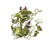
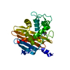
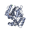
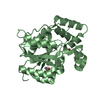
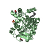
| Title | Structure of recombinant human DNase1L3 in complex with Mg2+ | ||||||
 Components Components | Deoxyribonuclease gamma | ||||||
 Keywords Keywords |  HYDROLASE / Nuclease / DNA hydrolase / Apoptosis / Lupus / Systemic Lupus Erythematosus / SLE / DNA clearance / Metal ion Nuclease / NET clearance HYDROLASE / Nuclease / DNA hydrolase / Apoptosis / Lupus / Systemic Lupus Erythematosus / SLE / DNA clearance / Metal ion Nuclease / NET clearance | ||||||
| Function / homology |  Function and homology information Function and homology information: / regulation of neutrophil mediated cytotoxicity / regulation of acute inflammatory response / DNA nuclease activity / programmed cell death involved in cell development / deoxyribonuclease I activity / apoptotic DNA fragmentation / neutrophil activation involved in immune response / Hydrolases; Acting on ester bonds; Endodeoxyribonucleases producing 5'-phosphomonoesters / DNA catabolic process ...: / regulation of neutrophil mediated cytotoxicity / regulation of acute inflammatory response / DNA nuclease activity / programmed cell death involved in cell development / deoxyribonuclease I activity / apoptotic DNA fragmentation / neutrophil activation involved in immune response / Hydrolases; Acting on ester bonds; Endodeoxyribonucleases producing 5'-phosphomonoesters / DNA catabolic process / DNA metabolic process / calcium ion binding / endoplasmic reticulum / DNA binding / extracellular region / nucleusSimilarity search - Function | ||||||
| Biological species |  Homo sapiens (human) Homo sapiens (human) | ||||||
| Method | X-RAY DIFFRACTION / SYNCHROTRON / MOLECULAR REPLACEMENT / Resolution: 2.22 Å | ||||||
 Authors Authors | McCord, J.J. / Keyel, P.A. / Sutton, R.B. | ||||||
 Citation Citation | Journal: Commun Biol / Year: 2022 Title: Structural features of Dnase1L3 responsible for serum antigen clearance. Authors: McCord, J.J. / Engavale, M. / Masoumzadeh, E. / Villarreal, J. / Mapp, B. / Latham, M.P. / Keyel, P.A. / Sutton, R.B. | ||||||
| History |
|
- Structure visualization
| Structure viewer | Molecule: MolmilJmol/JSmol |
|---|
- Downloads & links
-Download
| PDBx/mmCIF format | 7kiu.cif.gz | 475.4 KB | Display | PDBx/mmCIF format |
|---|---|---|---|---|
| PDB format | pdb7kiu.ent.gz | 375.7 KB | Display | PDB format |
| PDBx/mmJSON format | 7kiu.json.gz | Tree view | PDBx/mmJSON format | |
| Others |  Other downloads Other downloads |
-Validation report
| Arichive directory | https://data.pdbj.org/pub/pdb/validation_reports/ki/7kiuftp://data.pdbj.org/pub/pdb/validation_reports/ki/7kiu | HTTPS FTP |
|---|
-Related structure data
| Related structure data | 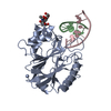 1dnkS S: Starting model for refinement |
|---|---|
| Similar structure data |


-Links
 PDBj
PDBj

- Assembly
| Deposited unit | 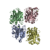
| ||||||||||||
|---|---|---|---|---|---|---|---|---|---|---|---|---|---|
| 1 | 
| ||||||||||||
| 2 | 
| ||||||||||||
| 3 | 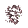
| ||||||||||||
| 4 | 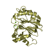
| ||||||||||||
| Unit cell |
|
-Components
| #1: Protein | Mass: 32772.387 Da / Num. of mol.: 4 Source method: isolated from a genetically manipulated source Source: (gene. exp.) Homo sapiens (human) / Gene: DNASE1L3, DHP2, DNAS1L3 / Plasmid: p202Details (production host): TEV cleavable MBP-His tagged fusion protein Production host:  Escherichia coli (E. coli) / Strain (production host): Rosetta-Gami 2(DE3) Escherichia coli (E. coli) / Strain (production host): Rosetta-Gami 2(DE3)References: UniProt: Q13609, Hydrolases; Acting on ester bonds; Endodeoxyribonucleases producing 5'-phosphomonoesters#2: Chemical | ChemComp-MG /   Mass: 24.305 Da / Num. of mol.: 12 / Source method: obtained synthetically / Formula: Mg Mass: 24.305 Da / Num. of mol.: 12 / Source method: obtained synthetically / Formula: Mg#3: Water | ChemComp-HOH / | Water Mass: 18.015 Da / Num. of mol.: 174 / Source method: isolated from a natural source / Formula: H2O Mass: 18.015 Da / Num. of mol.: 174 / Source method: isolated from a natural source / Formula: H2OHas ligand of interest | N | |
|---|
-Experimental details
-Experiment
| Experiment | Method: X-RAY DIFFRACTION / Number of used crystals: 1 |
|---|
- Sample preparation
| Crystal | Density Matthews: 2.17 Å3/Da / Density % sol: 43 % |
|---|---|
| Crystal grow | Temperature: 283 K / Method: vapor diffusion, hanging drop / pH: 8.5 Details: 19% PEG 8000, 250 mM Magnesium Chloride, 100 mM Tris pH 8.5 |
-Data collection
| Diffraction | Mean temperature: 100 K / Serial crystal experiment: N |
|---|---|
| Diffraction source | Source: SYNCHROTRON / Site: SSRL  / Beamline: BL14-1 / Wavelength: 1.12709 Å / Beamline: BL14-1 / Wavelength: 1.12709 Å |
| Detector | Type: RAYONIX MX325HE / Detector: CCD / Date: Dec 15, 2019 |
| Radiation | Monochromator: 1.12709 / Protocol: SINGLE WAVELENGTH / Monochromatic (M) / Laue (L): M / Scattering type: x-ray |
| Radiation wavelength | Wavelength: 1.12709 Å / Relative weight: 1 |
| Reflection | Resolution: 2.21→32.07 Å / Num. obs: 50424 / % possible obs: 94.9 % / Redundancy: 9.1 % / Biso Wilson estimate: 44.98 Å2 / CC1/2: 0.998 / Rmerge(I) obs: 0.145 / Rrim(I) all: 0.153 / Net I/σ(I): 8.5 |
| Reflection shell | Resolution: 2.21→2.25 Å / Redundancy: 3.9 % / Rmerge(I) obs: 1.391 / Mean I/σ(I) obs: 0.9 / Num. unique obs: 2368 / CC1/2: 0.491 / Rrim(I) all: 1.609 / % possible all: 90.3 |
- Processing
| Software |
| |||||||||||||||||||||||||||||||||||||||||||||||||||||||||||||||||||||||||||||||||||||||||||||||||||||||||||||||||||||||
|---|---|---|---|---|---|---|---|---|---|---|---|---|---|---|---|---|---|---|---|---|---|---|---|---|---|---|---|---|---|---|---|---|---|---|---|---|---|---|---|---|---|---|---|---|---|---|---|---|---|---|---|---|---|---|---|---|---|---|---|---|---|---|---|---|---|---|---|---|---|---|---|---|---|---|---|---|---|---|---|---|---|---|---|---|---|---|---|---|---|---|---|---|---|---|---|---|---|---|---|---|---|---|---|---|---|---|---|---|---|---|---|---|---|---|---|---|---|---|---|---|
| Refinement | Method to determine structure: MOLECULAR REPLACEMENT Starting model: 1DNK Resolution: 2.22→32.06 Å / SU ML: 0.4141 / Cross valid method: FREE R-VALUE / Phase error: 30.7442 Stereochemistry target values: GeoStd + Monomer Library + CDL v1.2
| |||||||||||||||||||||||||||||||||||||||||||||||||||||||||||||||||||||||||||||||||||||||||||||||||||||||||||||||||||||||
| Solvent computation | Shrinkage radii: 0.9 Å / VDW probe radii: 1.11 Å / Solvent model: FLAT BULK SOLVENT MODEL | |||||||||||||||||||||||||||||||||||||||||||||||||||||||||||||||||||||||||||||||||||||||||||||||||||||||||||||||||||||||
| Displacement parameters | Biso mean: 64.95 Å2 | |||||||||||||||||||||||||||||||||||||||||||||||||||||||||||||||||||||||||||||||||||||||||||||||||||||||||||||||||||||||
| Refinement step | Cycle: LAST / Resolution: 2.22→32.06 Å
| |||||||||||||||||||||||||||||||||||||||||||||||||||||||||||||||||||||||||||||||||||||||||||||||||||||||||||||||||||||||
| Refine LS restraints |
| |||||||||||||||||||||||||||||||||||||||||||||||||||||||||||||||||||||||||||||||||||||||||||||||||||||||||||||||||||||||
| LS refinement shell |
|
