 Movie
Movie Controller

[English] 日本語
 Yorodumi
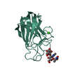
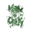
- PDB-7bll: Structure of SusD homologue BT3013 from Bacteroides thetaiotaomicron -
+ Open data

- Basic information
| Entry | Database: PDB / ID: 7bll | ||||||
|---|---|---|---|---|---|---|---|
| Title | Structure of SusD homologue BT3013 from Bacteroides thetaiotaomicron | ||||||
 Components Components | SusD homolog | ||||||
 Keywords Keywords | SUGAR BINDING PROTEIN /  Bacteroides thetaiotaomicron / surface glycan-binding protein (SusD homologue) / gut microbiome / mucins Bacteroides thetaiotaomicron / surface glycan-binding protein (SusD homologue) / gut microbiome / mucins | ||||||
| Function / homology | SusD-like, N-terminal / Starch-binding associating with outer membrane / RagB/SusD domain / SusD family / cell outer membrane / Tetratricopeptide-like helical domain superfamily / DI(HYDROXYETHYL)ETHER / SusD homolog Function and homology information Function and homology information | ||||||
| Biological species |  Bacteroides thetaiotaomicron (bacteria) Bacteroides thetaiotaomicron (bacteria) | ||||||
| Method | X-RAY DIFFRACTION / SYNCHROTRON / SAD / Resolution: 1.76 Å | ||||||
 Authors Authors | Costa, R.L. | ||||||
| Funding support |  Portugal, 1items Portugal, 1items
| ||||||
 Citation Citation | Journal: To Be Published Title: Structural basis for mucin-type O-glycan recognition by proteins of a Bacteroides thetaiotaomicron polysaccharide utilization loci Authors: Costa, R.L. / Correia, V.G. | ||||||
| History |
|
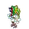
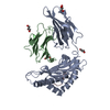
- Structure visualization
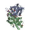
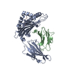
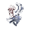
| Structure viewer | Molecule: MolmilJmol/JSmol |
|---|
- Downloads & links
-Download
| PDBx/mmCIF format | 7bll.cif.gz | 301.1 KB | Display | PDBx/mmCIF format |
|---|---|---|---|---|
| PDB format | pdb7bll.ent.gz | 207.1 KB | Display | PDB format |
| PDBx/mmJSON format | 7bll.json.gz | Tree view | PDBx/mmJSON format | |
| Others |  Other downloads Other downloads |
-Validation report
| Arichive directory | https://data.pdbj.org/pub/pdb/validation_reports/bl/7bllftp://data.pdbj.org/pub/pdb/validation_reports/bl/7bll | HTTPS FTP |
|---|
-Related structure data


-Links
 PDBj
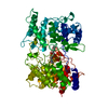
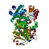
PDBj- Assembly
| Deposited unit | 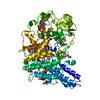
| ||||||||||||
|---|---|---|---|---|---|---|---|---|---|---|---|---|---|
| 1 |
| ||||||||||||
| Unit cell |
| ||||||||||||
| Components on special symmetry positions |
|
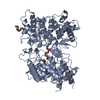
-Components
| #1: Protein | Mass: 73420.578 Da / Num. of mol.: 1 Source method: isolated from a genetically manipulated source Source: (gene. exp.) Bacteroides thetaiotaomicron (strain ATCC 29148 / DSM 2079 / NCTC 10582 / E50 / VPI-5482) (bacteria)Strain: ATCC 29148 / DSM 2079 / NCTC 10582 / E50 / VPI-5482 / Gene: BT_3013 Production host: Escherichia coli 'BL21-Gold(DE3)pLysS AG' (bacteria)References: UniProt: Q8A3E1 |
|---|---|
| #2: Chemical | ChemComp-PEG / Diethylene glycol  Mass: 106.120 Da / Num. of mol.: 1 / Source method: obtained synthetically / Formula: C4H10O3 / Feature type: SUBJECT OF INVESTIGATION Mass: 106.120 Da / Num. of mol.: 1 / Source method: obtained synthetically / Formula: C4H10O3 / Feature type: SUBJECT OF INVESTIGATION |
| #3: Chemical | ChemComp-EDO / Ethylene glycol  Mass: 62.068 Da / Num. of mol.: 1 / Source method: obtained synthetically / Formula: C2H6O2 / Feature type: SUBJECT OF INVESTIGATION Mass: 62.068 Da / Num. of mol.: 1 / Source method: obtained synthetically / Formula: C2H6O2 / Feature type: SUBJECT OF INVESTIGATION |
| #4: Water | ChemComp-HOH / Water Mass: 18.015 Da / Num. of mol.: 570 / Source method: isolated from a natural source / Formula: H2O Mass: 18.015 Da / Num. of mol.: 570 / Source method: isolated from a natural source / Formula: H2O |
| Has ligand of interest | Y |
-Experimental details
-Experiment
| Experiment | Method: X-RAY DIFFRACTION / Number of used crystals: 1 |
|---|
- Sample preparation
| Crystal | Density Matthews: 2.25 Å3/Da / Density % sol: 45.29 % |
|---|---|
| Crystal grow | Temperature: 293 K / Method: vapor diffusion, sitting drop Details: Protein at 14mg/ml 30% w/v PEG 4000, 0.1 M sodium citrate pH 5.5 and 0.2 M ammonium acetate |
-Data collection
| Diffraction | Mean temperature: 100 K / Serial crystal experiment: N |
|---|---|
| Diffraction source | Source: SYNCHROTRON / Site: ESRF  / Beamline: ID30B / Wavelength: 0.9794 Å / Beamline: ID30B / Wavelength: 0.9794 Å |
| Detector | Type: DECTRIS PILATUS3 S 6M / Detector: PIXEL / Date: Oct 2, 2018 |
| Radiation | Protocol: SINGLE WAVELENGTH / Monochromatic (M) / Laue (L): M / Scattering type: x-ray |
| Radiation wavelength | Wavelength: 0.9794 Å / Relative weight: 1 |
| Reflection | Resolution: 1.76→86.57 Å / Num. obs: 372154 / % possible obs: 99.4 % / Redundancy: 5.9 % / Biso Wilson estimate: 17.66 Å2 / CC1/2: 0.994 / Net I/σ(I): 15.5 |
| Reflection shell | Resolution: 1.76→1.8 Å / Num. unique obs: 19950 / CC1/2: 0.939 |
- Processing
| Software |
| |||||||||||||||||||||||||||||||||||||||||||||||||||||||||||||||||||||||||||||||||||||||||||||||||||||||||||||||||||||||||||||||||||||||||||||||||||||||||||||||||||||||||||||||||||||||||||||||||||||||||||||||||||||||||
|---|---|---|---|---|---|---|---|---|---|---|---|---|---|---|---|---|---|---|---|---|---|---|---|---|---|---|---|---|---|---|---|---|---|---|---|---|---|---|---|---|---|---|---|---|---|---|---|---|---|---|---|---|---|---|---|---|---|---|---|---|---|---|---|---|---|---|---|---|---|---|---|---|---|---|---|---|---|---|---|---|---|---|---|---|---|---|---|---|---|---|---|---|---|---|---|---|---|---|---|---|---|---|---|---|---|---|---|---|---|---|---|---|---|---|---|---|---|---|---|---|---|---|---|---|---|---|---|---|---|---|---|---|---|---|---|---|---|---|---|---|---|---|---|---|---|---|---|---|---|---|---|---|---|---|---|---|---|---|---|---|---|---|---|---|---|---|---|---|---|---|---|---|---|---|---|---|---|---|---|---|---|---|---|---|---|---|---|---|---|---|---|---|---|---|---|---|---|---|---|---|---|---|---|---|---|---|---|---|---|---|---|---|---|---|---|---|---|---|
| Refinement | Method to determine structure: SAD / Resolution: 1.76→56.97 Å / SU ML: 0.2002 / Cross valid method: FREE R-VALUE / σ(F): 1.97 / Phase error: 20.8474 Stereochemistry target values: GeoStd + Monomer Library + CDL v1.2
| |||||||||||||||||||||||||||||||||||||||||||||||||||||||||||||||||||||||||||||||||||||||||||||||||||||||||||||||||||||||||||||||||||||||||||||||||||||||||||||||||||||||||||||||||||||||||||||||||||||||||||||||||||||||||
| Solvent computation | Shrinkage radii: 0.9 Å / VDW probe radii: 1.11 Å / Solvent model: FLAT BULK SOLVENT MODEL | |||||||||||||||||||||||||||||||||||||||||||||||||||||||||||||||||||||||||||||||||||||||||||||||||||||||||||||||||||||||||||||||||||||||||||||||||||||||||||||||||||||||||||||||||||||||||||||||||||||||||||||||||||||||||
| Displacement parameters | Biso mean: 20.61 Å2 | |||||||||||||||||||||||||||||||||||||||||||||||||||||||||||||||||||||||||||||||||||||||||||||||||||||||||||||||||||||||||||||||||||||||||||||||||||||||||||||||||||||||||||||||||||||||||||||||||||||||||||||||||||||||||
| Refinement step | Cycle: LAST / Resolution: 1.76→56.97 Å
| |||||||||||||||||||||||||||||||||||||||||||||||||||||||||||||||||||||||||||||||||||||||||||||||||||||||||||||||||||||||||||||||||||||||||||||||||||||||||||||||||||||||||||||||||||||||||||||||||||||||||||||||||||||||||
| Refine LS restraints |
| |||||||||||||||||||||||||||||||||||||||||||||||||||||||||||||||||||||||||||||||||||||||||||||||||||||||||||||||||||||||||||||||||||||||||||||||||||||||||||||||||||||||||||||||||||||||||||||||||||||||||||||||||||||||||
| LS refinement shell |
|
