 Movie
Movie Controller


+ Open data

- Basic information
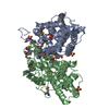
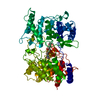
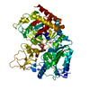
| Entry | Database: PDB / ID: 3sqc | ||||||
|---|---|---|---|---|---|---|---|
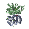
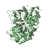
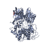
| Title | SQUALENE-HOPENE CYCLASE | ||||||
 Components Components | SQUALENE--HOPENE CYCLASE | ||||||
 Keywords Keywords |  ISOMERASE / TRITERPENE CYCLASE / MONOTOPIC MEMBRANE PROTEIN / QW-SEQUENCE REPEAT / CHOLESTEROL BIOSYNTHESIS ISOMERASE / TRITERPENE CYCLASE / MONOTOPIC MEMBRANE PROTEIN / QW-SEQUENCE REPEAT / CHOLESTEROL BIOSYNTHESIS | ||||||
| Function / homology |  Function and homology information Function and homology informationsqualene-hopanol cyclase / squalene-hopene cyclase / squalene-hopene cyclase activity / lanosterol synthase activity / triterpenoid biosynthetic process / ergosterol biosynthetic process / cholesterol biosynthetic process / lipid droplet / lyase activity / plasma membraneSimilarity search - Function | ||||||
| Biological species |  Alicyclobacillus acidocaldarius (bacteria) Alicyclobacillus acidocaldarius (bacteria) | ||||||
| Method | X-RAY DIFFRACTION / MOLECULAR REPLACEMENT / Resolution: 2.8 Å | ||||||
 Authors Authors | Wendt, K.U. / Schulz, G.E. | ||||||
 Citation Citation | Journal: J.Mol.Biol. / Year: 1999 Title: The structure of the membrane protein squalene-hopene cyclase at 2.0 A resolution. Authors: Wendt, K.U. / Lenhart, A. / Schulz, G.E. #1: Journal: Science / Year: 1997Title: Structure and Function of a Squalene Cyclase Authors: Wendt, K.U. / Poralla, K. / Schulz, G.E. #2: Journal: Protein Sci. / Year: 1997Title: Crystallization and Preliminary X-Ray Crystallographic Analysis of Squalene-Hopene Cyclase from Alicyclobacillus Acidocaldarius Authors: Wendt, K.U. / Feil, C. / Lenhart, A. / Poralla, K. / Schulz, G.E. | ||||||
| History |
|
- Structure visualization
| Structure viewer | Molecule: MolmilJmol/JSmol |
|---|
- Downloads & links
-Download
| PDBx/mmCIF format | 3sqc.cif.gz | 362.9 KB | Display | PDBx/mmCIF format |
|---|---|---|---|---|
| PDB format | pdb3sqc.ent.gz | 301.1 KB | Display | PDB format |
| PDBx/mmJSON format | 3sqc.json.gz | Tree view | PDBx/mmJSON format | |
| Others |  Other downloads Other downloads |
-Validation report
| Arichive directory | https://data.pdbj.org/pub/pdb/validation_reports/sq/3sqcftp://data.pdbj.org/pub/pdb/validation_reports/sq/3sqc | HTTPS FTP |
|---|
-Related structure data
| Related structure data |  2sqcC  1sqcS S: Starting model for refinement C: citing same article ( |
|---|---|
| Similar structure data |


-Links
 PDBj
PDBj

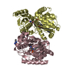
- Assembly
| Deposited unit | 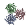
| ||||||||||||||||
|---|---|---|---|---|---|---|---|---|---|---|---|---|---|---|---|---|---|
| 1 | 
| ||||||||||||||||
| 2 | 
| ||||||||||||||||
| 3 | 
| ||||||||||||||||
| Unit cell |
| ||||||||||||||||
| Noncrystallographic symmetry (NCS) | NCS oper:
|
-Components
| #1: Protein | Mass: 71638.094 Da / Num. of mol.: 3 / Mutation: D376C Source method: isolated from a genetically manipulated source Source: (gene. exp.) Alicyclobacillus acidocaldarius (bacteria)Description: THERMOSTABLE, ACIDOPHILIC / Cell line: JM105 / Cellular location: MEMBRANE Biological membrane / Plasmid: PKK223-3 / Species (production host): Escherichia coli / Cell line (production host): JM105 / Cellular location (production host): CYTOPLASMIC MEMBRANE / Production host: Escherichia coli K12 (bacteria) / Strain (production host): K12References: UniProt: P33247, Isomerases; Intramolecular transferases; Transferring other groups#2: Water | ChemComp-HOH / | Water Mass: 18.015 Da / Num. of mol.: 114 / Source method: isolated from a natural source / Formula: H2O Mass: 18.015 Da / Num. of mol.: 114 / Source method: isolated from a natural source / Formula: H2O |
|---|
-Experimental details
-Experiment
| Experiment | Method: X-RAY DIFFRACTION / Number of used crystals: 3 |
|---|
- Sample preparation
| Crystal | Density Matthews: 3.3 Å3/Da / Density % sol: 63 % | ||||||||||||||||||||||||||||||
|---|---|---|---|---|---|---|---|---|---|---|---|---|---|---|---|---|---|---|---|---|---|---|---|---|---|---|---|---|---|---|---|
| Crystal grow | pH: 4.8 / Details: pH 4.8 | ||||||||||||||||||||||||||||||
| Crystal grow | *PLUS Method: vapor diffusion, hanging drop / Details: Wendt, K.U., (1997) Protein Sci., 6, 722. | ||||||||||||||||||||||||||||||
| Components of the solutions | *PLUS
|
-Data collection
| Diffraction | Mean temperature: 298 K |
|---|---|
| Diffraction source | Source: ROTATING ANODE / Type: RIGAKU RUH2R / Wavelength: 1.5418 |
| Detector | Type: SIEMENS / Detector: AREA DETECTOR |
| Radiation | Monochromator: GRAPHITE(002) / Monochromatic (M) / Laue (L): M / Scattering type: x-ray |
| Radiation wavelength | Wavelength: 1.5418 Å / Relative weight: 1 |
| Reflection | Resolution: 2.8→40 Å / Num. obs: 57421 / % possible obs: 83 % / Redundancy: 3.9 % / Rsym value: 0.047 / Net I/σ(I): 5.9 |
| Reflection shell | Resolution: 2.8→2.9 Å / Redundancy: 1.4 % / Mean I/σ(I) obs: 4.1 / Rsym value: 0.17 / % possible all: 44 |
- Processing
| Software |
| ||||||||||||||||||||||||||||||||||||||||||||||||||||||||||||
|---|---|---|---|---|---|---|---|---|---|---|---|---|---|---|---|---|---|---|---|---|---|---|---|---|---|---|---|---|---|---|---|---|---|---|---|---|---|---|---|---|---|---|---|---|---|---|---|---|---|---|---|---|---|---|---|---|---|---|---|---|---|
| Refinement | Method to determine structure: MOLECULAR REPLACEMENT Starting model: 1SQC Resolution: 2.8→20 Å / Data cutoff high absF: 1000000 / Data cutoff low absF: 0.001 / Cross valid method: THROUGHOUT / σ(F): 0
| ||||||||||||||||||||||||||||||||||||||||||||||||||||||||||||
| Displacement parameters | Biso mean: 50 Å2 | ||||||||||||||||||||||||||||||||||||||||||||||||||||||||||||
| Refine analyze | Luzzati coordinate error obs: 0.45 Å / Luzzati d res low obs: 4.6 Å / Luzzati sigma a obs: 0.5 Å | ||||||||||||||||||||||||||||||||||||||||||||||||||||||||||||
| Refinement step | Cycle: LAST / Resolution: 2.8→20 Å
| ||||||||||||||||||||||||||||||||||||||||||||||||||||||||||||
| Refine LS restraints |
| ||||||||||||||||||||||||||||||||||||||||||||||||||||||||||||
| Refine LS restraints NCS | NCS model details: RESTRAINTS | ||||||||||||||||||||||||||||||||||||||||||||||||||||||||||||
| LS refinement shell | Resolution: 2.8→2.93 Å
| ||||||||||||||||||||||||||||||||||||||||||||||||||||||||||||
| Software | *PLUS Name: X-PLOR / Version: 3.8 / Classification: refinement | ||||||||||||||||||||||||||||||||||||||||||||||||||||||||||||
| Refine LS restraints | *PLUS
|
