 Movie
Movie Controller

[English] 日本語
 Yorodumi
- PDB-3bjx: Structure of a Group I haloacid dehalogenase from Pseudomonas put... -
+ Open data

- Basic information
| Entry | Database: PDB / ID: 3bjx | ||||||
|---|---|---|---|---|---|---|---|
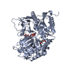
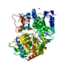
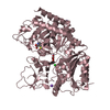
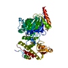
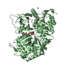
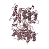
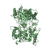
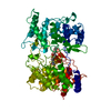
| Title | Structure of a Group I haloacid dehalogenase from Pseudomonas putida strain PP3 | ||||||
 Components Components | Halocarboxylic acid dehalogenase DehI | ||||||
 Keywords Keywords |  HYDROLASE / Plasmid HYDROLASE / Plasmid | ||||||
| Function / homology | 2-haloacid dehalogenase, DehI / Halocarboxylic acid dehydrogenase DehI / hydrolase activity, acting on acid halide bonds, in C-halide compounds / Halocarboxylic acid dehalogenase DehI Function and homology information Function and homology information | ||||||
| Biological species |  Pseudomonas putida (bacteria) Pseudomonas putida (bacteria) | ||||||
| Method | X-RAY DIFFRACTION / SYNCHROTRON / MAD / Resolution: 2.3 Å | ||||||
 Authors Authors | Schmidberger, J.W. / Wilce, M.C.J. | ||||||
 Citation Citation | Journal: J.Mol.Biol. / Year: 2008 Title: The crystal structure of DehI reveals a new alpha-haloacid dehalogenase fold and active-site mechanism Authors: Schmidberger, J.W. / Wilce, J.A. / Weightman, A.J. / Whisstock, J.C. / Wilce, M.C.J. | ||||||
| History |
| ||||||
| Remark 40 | MOLPROBITY STRUCTURE VALIDATION PROGRAMS : MOLPROBITY (KING, REDUCE, AND PROBE) AUTHORS : I.W. ...MOLPROBITY STRUCTURE VALIDATION PROGRAMS : MOLPROBITY (KING, REDUCE, AND PROBE) AUTHORS : I.W.DAVIS,V.B.CHEN, : R.M.IMMORMINO,J.J.HEADD,W.B.ARENDALL,J.M.WORD URL : HTTP://KINEMAGE.BIOCHEM.DUKE.EDU/MOLPROBITY/ AUTHORS : I.W.DAVIS,A.LEAVER-FAY,V.B.CHEN,J.N.BLOCK, : G.J.KAPRAL,X.WANG,L.W.MURRAY,W.B.ARENDALL, : J.SNOEYINK,J.S.RICHARDSON,D.C.RICHARDSON REFERENCE : MOLPROBITY: ALL-ATOM CONTACTS AND STRUCTURE : VALIDATION FOR PROTEINS AND NUCLEIC ACIDS : NUCLEIC ACIDS RESEARCH. 2007;35:W375-83. MOLPROBITY OUTPUT SCORES: ALL-ATOM CLASHSCORE : 12.67 BAD ROTAMERS : 2.9% 29/1006 (TARGET 0-1%) RAMACHANDRAN OUTLIERS : 0.5% 6/1203 (TARGET 0.2%) RAMACHANDRAN FAVORED : 98.3% 1183/1203 (TARGET 98.0%) |
- Structure visualization
| Structure viewer | Molecule: MolmilJmol/JSmol |
|---|
- Downloads & links
-Download
| PDBx/mmCIF format | 3bjx.cif.gz | 247.8 KB | Display | PDBx/mmCIF format |
|---|---|---|---|---|
| PDB format | pdb3bjx.ent.gz | 208.9 KB | Display | PDB format |
| PDBx/mmJSON format | 3bjx.json.gz | Tree view | PDBx/mmJSON format | |
| Others |  Other downloads Other downloads |
-Validation report
| Arichive directory | https://data.pdbj.org/pub/pdb/validation_reports/bj/3bjxftp://data.pdbj.org/pub/pdb/validation_reports/bj/3bjx | HTTPS FTP |
|---|
-Related structure data
| Similar structure data |
|---|


-Links
 PDBj
PDBj- Assembly
| Deposited unit | 
| ||||||||
|---|---|---|---|---|---|---|---|---|---|
| 1 | 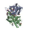
| ||||||||
| 2 | 
| ||||||||
| Unit cell |
|
-Components
| #1: Protein | Mass: 34866.633 Da / Num. of mol.: 4 Source method: isolated from a genetically manipulated source Source: (gene. exp.) Pseudomonas putida (bacteria) / Strain: PP3 / Gene: dehI / Plasmid: pET15b / Production host: Escherichia coli (E. coli) / Strain (production host): Nova Blue / References: UniProt: Q8GJ84, (S)-2-haloacid dehalogenase#2: Chemical | ChemComp-SO4 / Sulfate  Mass: 96.063 Da / Num. of mol.: 16 / Source method: obtained synthetically / Formula: SO4 Mass: 96.063 Da / Num. of mol.: 16 / Source method: obtained synthetically / Formula: SO4#3: Water | ChemComp-HOH / | Water Mass: 18.015 Da / Num. of mol.: 449 / Source method: isolated from a natural source / Formula: H2O Mass: 18.015 Da / Num. of mol.: 449 / Source method: isolated from a natural source / Formula: H2OSequence details | THE PUBLISHED SEQUENCE FOR DEHI WITH ASSESSION CODE Q8GJ84 IS WRONG FOR RESIDUE 229. A PROLINE IS ...THE PUBLISHED SEQUENCE FOR DEHI WITH ASSESSION CODE Q8GJ84 IS WRONG FOR RESIDUE 229. A PROLINE IS REPORTED FOR THIS RESIDUE BUT IS IN FACT AN ARGININE AS REPORTED IN THIS STRUCTURE. | |
|---|
-Experimental details
-Experiment
| Experiment | Method: X-RAY DIFFRACTION / Number of used crystals: 1 |
|---|
- Sample preparation
| Crystal | Density Matthews: 2.24 Å3/Da / Density % sol: 40.17 % |
|---|---|
| Crystal grow | Temperature: 298 K / Method: hanging drop / pH: 6 Details: 25% PEG 3350 w/v, 0.4M Li2SO4, 0.1M Bis-Tris pH 6.0, hanging drop, temperature 298K |
-Data collection
| Diffraction |
| ||||||||||||||||||||||||||||||||||||||||||||||||||||||||||||||||||||||||||||||||||||||||
|---|---|---|---|---|---|---|---|---|---|---|---|---|---|---|---|---|---|---|---|---|---|---|---|---|---|---|---|---|---|---|---|---|---|---|---|---|---|---|---|---|---|---|---|---|---|---|---|---|---|---|---|---|---|---|---|---|---|---|---|---|---|---|---|---|---|---|---|---|---|---|---|---|---|---|---|---|---|---|---|---|---|---|---|---|---|---|---|---|---|
| Diffraction source |
| ||||||||||||||||||||||||||||||||||||||||||||||||||||||||||||||||||||||||||||||||||||||||
| Detector |
| ||||||||||||||||||||||||||||||||||||||||||||||||||||||||||||||||||||||||||||||||||||||||
| Radiation |
| ||||||||||||||||||||||||||||||||||||||||||||||||||||||||||||||||||||||||||||||||||||||||
| Radiation wavelength |
| ||||||||||||||||||||||||||||||||||||||||||||||||||||||||||||||||||||||||||||||||||||||||
| Reflection | Resolution: 2.3→75.024 Å / Num. obs: 48240 / % possible obs: 96.3 % / Redundancy: 2.5 % / Rmerge(I) obs: 0.118 / Rsym value: 0.118 / Net I/σ(I): 6.1 | ||||||||||||||||||||||||||||||||||||||||||||||||||||||||||||||||||||||||||||||||||||||||
| Reflection shell |
|

-Phasing
| Phasing | Method: MAD | ||||||||||||||||||||||||||||||||||||||||||||||||||||||||||||||||||||||||||||||||||||||||||||||||||||||||||||||||||||||||||||||||||||||||||||||||||||||||||||||||||||||||||||||||||||||||||||||||||||||||||||||||||||||||||||||||
|---|---|---|---|---|---|---|---|---|---|---|---|---|---|---|---|---|---|---|---|---|---|---|---|---|---|---|---|---|---|---|---|---|---|---|---|---|---|---|---|---|---|---|---|---|---|---|---|---|---|---|---|---|---|---|---|---|---|---|---|---|---|---|---|---|---|---|---|---|---|---|---|---|---|---|---|---|---|---|---|---|---|---|---|---|---|---|---|---|---|---|---|---|---|---|---|---|---|---|---|---|---|---|---|---|---|---|---|---|---|---|---|---|---|---|---|---|---|---|---|---|---|---|---|---|---|---|---|---|---|---|---|---|---|---|---|---|---|---|---|---|---|---|---|---|---|---|---|---|---|---|---|---|---|---|---|---|---|---|---|---|---|---|---|---|---|---|---|---|---|---|---|---|---|---|---|---|---|---|---|---|---|---|---|---|---|---|---|---|---|---|---|---|---|---|---|---|---|---|---|---|---|---|---|---|---|---|---|---|---|---|---|---|---|---|---|---|---|---|---|---|---|---|---|---|---|
| Phasing set |
| ||||||||||||||||||||||||||||||||||||||||||||||||||||||||||||||||||||||||||||||||||||||||||||||||||||||||||||||||||||||||||||||||||||||||||||||||||||||||||||||||||||||||||||||||||||||||||||||||||||||||||||||||||||||||||||||||
| Phasing MAD set |
| ||||||||||||||||||||||||||||||||||||||||||||||||||||||||||||||||||||||||||||||||||||||||||||||||||||||||||||||||||||||||||||||||||||||||||||||||||||||||||||||||||||||||||||||||||||||||||||||||||||||||||||||||||||||||||||||||
| Phasing MAD set site |
| ||||||||||||||||||||||||||||||||||||||||||||||||||||||||||||||||||||||||||||||||||||||||||||||||||||||||||||||||||||||||||||||||||||||||||||||||||||||||||||||||||||||||||||||||||||||||||||||||||||||||||||||||||||||||||||||||
| Phasing dm | FOM : 0.65 / FOM acentric: 0.66 / FOM centric: 0.6 / Reflection: 27694 / Reflection acentric: 26674 / Reflection centric: 1020 | ||||||||||||||||||||||||||||||||||||||||||||||||||||||||||||||||||||||||||||||||||||||||||||||||||||||||||||||||||||||||||||||||||||||||||||||||||||||||||||||||||||||||||||||||||||||||||||||||||||||||||||||||||||||||||||||||
| Phasing dm shell |
|
- Processing
| Software |
| |||||||||||||||||||||||||||||||||||||||||||||||||||||||||||||||||||||||||||||||||||||||||||||||||||||||||||||||||||||||||||||||||||||||||||||||||||||||||||||||||||||
|---|---|---|---|---|---|---|---|---|---|---|---|---|---|---|---|---|---|---|---|---|---|---|---|---|---|---|---|---|---|---|---|---|---|---|---|---|---|---|---|---|---|---|---|---|---|---|---|---|---|---|---|---|---|---|---|---|---|---|---|---|---|---|---|---|---|---|---|---|---|---|---|---|---|---|---|---|---|---|---|---|---|---|---|---|---|---|---|---|---|---|---|---|---|---|---|---|---|---|---|---|---|---|---|---|---|---|---|---|---|---|---|---|---|---|---|---|---|---|---|---|---|---|---|---|---|---|---|---|---|---|---|---|---|---|---|---|---|---|---|---|---|---|---|---|---|---|---|---|---|---|---|---|---|---|---|---|---|---|---|---|---|---|---|---|---|---|
| Refinement | Method to determine structure: MAD / Resolution: 2.3→38.15 Å / Cor.coef. Fo:Fc: 0.942 / Cor.coef. Fo:Fc free: 0.896 / SU B: 7.727 / SU ML: 0.188 / Cross valid method: THROUGHOUT / σ(F): 0 / ESU R: 0.559 / ESU R Free: 0.277 / Stereochemistry target values: MAXIMUM LIKELIHOOD / Details: HYDROGENS HAVE BEEN ADDED IN THE RIDING POSITIONS
| |||||||||||||||||||||||||||||||||||||||||||||||||||||||||||||||||||||||||||||||||||||||||||||||||||||||||||||||||||||||||||||||||||||||||||||||||||||||||||||||||||||
| Solvent computation | Ion probe radii: 0.8 Å / Shrinkage radii: 0.8 Å / VDW probe radii: 1.2 Å / Solvent model: MASK | |||||||||||||||||||||||||||||||||||||||||||||||||||||||||||||||||||||||||||||||||||||||||||||||||||||||||||||||||||||||||||||||||||||||||||||||||||||||||||||||||||||
| Displacement parameters | Biso mean: 24.182 Å2
| |||||||||||||||||||||||||||||||||||||||||||||||||||||||||||||||||||||||||||||||||||||||||||||||||||||||||||||||||||||||||||||||||||||||||||||||||||||||||||||||||||||
| Refinement step | Cycle: LAST / Resolution: 2.3→38.15 Å
| |||||||||||||||||||||||||||||||||||||||||||||||||||||||||||||||||||||||||||||||||||||||||||||||||||||||||||||||||||||||||||||||||||||||||||||||||||||||||||||||||||||
| Refine LS restraints |
| |||||||||||||||||||||||||||||||||||||||||||||||||||||||||||||||||||||||||||||||||||||||||||||||||||||||||||||||||||||||||||||||||||||||||||||||||||||||||||||||||||||
| LS refinement shell | Resolution: 2.3→2.36 Å / Total num. of bins used: 20
|
