 Movie
Movie Controller

[English] 日本語
 Yorodumi
- PDB-7blh: Structure of CBM BT3015C from Bacteroides thetaiotaomicron in com... -
+ Open data

- Basic information
| Entry | Database: PDB / ID: 7blh | ||||||
|---|---|---|---|---|---|---|---|
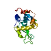
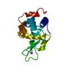
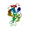
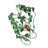
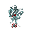
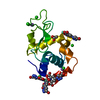
| Title | Structure of CBM BT3015C from Bacteroides thetaiotaomicron in complex with O-GalNAc core 1-Thr | ||||||
 Components Components | family 32 carbohydrate-binding module from Bacteroides thetaiotaomicron | ||||||
 Keywords Keywords | SUGAR BINDING PROTEIN /  Bacteroides thetaiotaomicron / carbohydrate-binding module / gut microbiome / mucins / O-GalNAc core 1-Thr Bacteroides thetaiotaomicron / carbohydrate-binding module / gut microbiome / mucins / O-GalNAc core 1-Thr | ||||||
| Function / homology |  Function and homology information Function and homology informationViral BACON domain / Bacteroidetes-Associated Carbohydrate-binding Often N-terminal / Peptidase family M60 domain / Peptidase M60, enhancin-like domain 3 / Peptidase M60, enhancin and enhancin-like / Peptidase family M60 domain profile. / Peptidase M60-like family / Coagulation factors 5/8 type C domain (FA58C) profile. / F5/8 type C domain / Coagulation factor 5/8 C-terminal domain ...Viral BACON domain / Bacteroidetes-Associated Carbohydrate-binding Often N-terminal / Peptidase family M60 domain / Peptidase M60, enhancin-like domain 3 / Peptidase M60, enhancin and enhancin-like / Peptidase family M60 domain profile. / Peptidase M60-like family / Coagulation factors 5/8 type C domain (FA58C) profile. / F5/8 type C domain / Coagulation factor 5/8 C-terminal domain / Galactose-binding-like domain superfamily / Prokaryotic membrane lipoprotein lipid attachment site profile. / Immunoglobulin-like foldSimilarity search - Domain/homology | ||||||
| Biological species |  Bacteroides thetaiotaomicron VPI-5482 (bacteria) Bacteroides thetaiotaomicron VPI-5482 (bacteria) | ||||||
| Method | X-RAY DIFFRACTION / MOLECULAR REPLACEMENT / Resolution: 1.79 Å | ||||||
 Authors Authors | Costa, R.L. / Carvalho, A.L. | ||||||
| Funding support |  Portugal, 1items Portugal, 1items
| ||||||
 Citation Citation | Journal: To Be Published Title: Structural basis for mucin-type O-glycan recognition by proteins of a Bacteroides thetaiotaomicron polysaccharide utilization loci Authors: Costa, R.L. / Correia, V.G. | ||||||
| History |
|
- Structure visualization
| Structure viewer | Molecule: MolmilJmol/JSmol |
|---|
- Downloads & links
-Download
| PDBx/mmCIF format | 7blh.cif.gz | 59.7 KB | Display | PDBx/mmCIF format |
|---|---|---|---|---|
| PDB format | pdb7blh.ent.gz | Display | PDB format | |
| PDBx/mmJSON format | 7blh.json.gz | Tree view | PDBx/mmJSON format | |
| Others |  Other downloads Other downloads |
-Validation report
| Arichive directory | https://data.pdbj.org/pub/pdb/validation_reports/bl/7blhftp://data.pdbj.org/pub/pdb/validation_reports/bl/7blh | HTTPS FTP |
|---|
-Related structure data
| Related structure data |  7blgSC  7bljC  7blkC  7bllC S: Starting model for refinement C: citing same article ( |
|---|---|
| Similar structure data |


-Links
 PDBj
PDBj


- Assembly
| Deposited unit | 
| ||||||||||||
|---|---|---|---|---|---|---|---|---|---|---|---|---|---|
| 1 |
| ||||||||||||
| Unit cell |
|
-Components
| #1: Protein | Mass: 17917.562 Da / Num. of mol.: 1 Source method: isolated from a genetically manipulated source Source: (gene. exp.) Bacteroides thetaiotaomicron VPI-5482 (bacteria)Gene: BT_3015 Production host: Escherichia coli 'BL21-Gold(DE3)pLysS AG' (bacteria)References: UniProt: Q8A3D9 |
|---|---|
| #2: Polysaccharide | beta-D-galactopyranose-(1-3)-2-acetamido-2-deoxy-alpha-D-galactopyranose / Mass: 383.349 Da / Num. of mol.: 1 Source method: isolated from a genetically manipulated source |
| #3: Chemical | ChemComp-THR / Threonine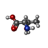  Type: L-peptide linking / Mass: 119.119 Da / Num. of mol.: 1 / Source method: obtained synthetically / Formula: C4H9NO3 / Feature type: SUBJECT OF INVESTIGATION Type: L-peptide linking / Mass: 119.119 Da / Num. of mol.: 1 / Source method: obtained synthetically / Formula: C4H9NO3 / Feature type: SUBJECT OF INVESTIGATION |
| #4: Chemical | ChemComp-CA /   Mass: 40.078 Da / Num. of mol.: 1 / Source method: obtained synthetically / Formula: Ca Mass: 40.078 Da / Num. of mol.: 1 / Source method: obtained synthetically / Formula: Ca |
| #5: Water | ChemComp-HOH / Water Mass: 18.015 Da / Num. of mol.: 231 / Source method: isolated from a natural source / Formula: H2O Mass: 18.015 Da / Num. of mol.: 231 / Source method: isolated from a natural source / Formula: H2O |
| Has ligand of interest | Y |
-Experimental details
-Experiment
| Experiment | Method: X-RAY DIFFRACTION / Number of used crystals: 1 |
|---|
- Sample preparation
| Crystal | Density Matthews: 2.23 Å3/Da / Density % sol: 44.79 % |
|---|---|
| Crystal grow | Temperature: 293 K / Method: vapor diffusion, sitting drop Details: Protein at 8mg/ml 0.2 M potassium formate and 20 % w/v PEG 6000 |
-Data collection
| Diffraction | Mean temperature: 110 K / Serial crystal experiment: N |
|---|---|
| Diffraction source | Source: SEALED TUBE / Type: BRUKER IMUS MICROFOCUS / Wavelength: 1.5418 Å |
| Detector | Type: BRUKER PHOTON 100 / Detector: CMOS / Date: Apr 17, 2019 |
| Radiation | Protocol: SINGLE WAVELENGTH / Monochromatic (M) / Laue (L): M / Scattering type: x-ray |
| Radiation wavelength | Wavelength: 1.5418 Å / Relative weight: 1 |
| Reflection | Resolution: 1.79→23.65 Å / Num. obs: 15596 / % possible obs: 99.2 % / Redundancy: 5.82 % / Biso Wilson estimate: 13.5 Å2 / Rmerge(I) obs: 0.092 / Net I/σ(I): 19.09 |
| Reflection shell | Resolution: 1.79→1.89 Å / Rmerge(I) obs: 0.256 / Num. unique obs: 2194 |
- Processing
| Software |
| ||||||||||||||||||||||||||||||||||||||||||
|---|---|---|---|---|---|---|---|---|---|---|---|---|---|---|---|---|---|---|---|---|---|---|---|---|---|---|---|---|---|---|---|---|---|---|---|---|---|---|---|---|---|---|---|
| Refinement | Method to determine structure: MOLECULAR REPLACEMENT Starting model: 7BLG Resolution: 1.79→20.42 Å / SU ML: 0.1465 / Cross valid method: FREE R-VALUE / σ(F): 1.41 / Phase error: 18.283 Stereochemistry target values: GeoStd + Monomer Library + CDL v1.2
| ||||||||||||||||||||||||||||||||||||||||||
| Solvent computation | Shrinkage radii: 0.9 Å / VDW probe radii: 1.11 Å / Solvent model: FLAT BULK SOLVENT MODEL | ||||||||||||||||||||||||||||||||||||||||||
| Displacement parameters | Biso mean: 9.92 Å2 | ||||||||||||||||||||||||||||||||||||||||||
| Refinement step | Cycle: LAST / Resolution: 1.79→20.42 Å
| ||||||||||||||||||||||||||||||||||||||||||
| Refine LS restraints |
| ||||||||||||||||||||||||||||||||||||||||||
| LS refinement shell |
|
