 Movie
Movie Controller

[English] 日本語
 Yorodumi
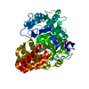
- PDB-7bj1: Crystal structure of SMYD3 with diperodon S enantiomer bound to a... -
+ Open data

- Basic information
| Entry | Database: PDB / ID: 7bj1 | |||||||||
|---|---|---|---|---|---|---|---|---|---|---|
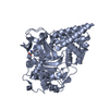
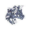
| Title | Crystal structure of SMYD3 with diperodon S enantiomer bound to allosteric site | |||||||||
 Components Components | Histone-lysine N-methyltransferase SMYD3 | |||||||||
 Keywords Keywords |  ONCOPROTEIN / Methyltransferase / complex / inhibitor ONCOPROTEIN / Methyltransferase / complex / inhibitor | |||||||||
| Function / homology |  Function and homology information Function and homology informationhistone H3K36 dimethyltransferase activity / histone H4 methyltransferase activity / [histone H3]-lysine4 N-trimethyltransferase / myotube cell development / histone H3K4 trimethyltransferase activity / RNA polymerase II intronic transcription regulatory region sequence-specific DNA binding / RNA polymerase II complex binding / cellular response to dexamethasone stimulus / establishment of protein localization / PKMTs methylate histone lysines ...histone H3K36 dimethyltransferase activity / histone H4 methyltransferase activity / [histone H3]-lysine4 N-trimethyltransferase / myotube cell development / histone H3K4 trimethyltransferase activity / RNA polymerase II intronic transcription regulatory region sequence-specific DNA binding / RNA polymerase II complex binding / cellular response to dexamethasone stimulus / establishment of protein localization / PKMTs methylate histone lysines / nucleosome assembly / positive regulation of peptidyl-serine phosphorylation / methylation / RNA polymerase II cis-regulatory region sequence-specific DNA binding / positive regulation of transcription by RNA polymerase II / nucleoplasm / metal ion binding / nucleus / cytosolSimilarity search - Function | |||||||||
| Biological species |  Homo sapiens (human) Homo sapiens (human) | |||||||||
| Method | X-RAY DIFFRACTION / SYNCHROTRON / MOLECULAR REPLACEMENT / Resolution: 1.61 Å | |||||||||
 Authors Authors | Talibov, V.O. / Cederfelt, D. / Dobritzsch, D. / Danielson, U.H. | |||||||||
| Funding support |  Sweden, Sweden,  Italy, 2items Italy, 2items
| |||||||||
 Citation Citation | Journal: Chembiochem / Year: 2021 Title: Discovery of an Allosteric Ligand Binding Site in SMYD3 Lysine Methyltransferase Authors: Talibov, V.O. / Fabini, E. / FitzGerald, E.A. / Tedesco, D. / Cederfeldt, D. / Talu, M.J. / Rachman, M.M. / Mihalic, F. / Manoni, E. / Naldi, M. / Sanese, P. / Forte, G. / Barril, X. / ...Authors: Talibov, V.O. / Fabini, E. / FitzGerald, E.A. / Tedesco, D. / Cederfeldt, D. / Talu, M.J. / Rachman, M.M. / Mihalic, F. / Manoni, E. / Naldi, M. / Sanese, P. / Forte, G. / Barril, X. / Simone, C. / Bartolini, M. / Dobritzsch, D. / Danielson, U.H. | |||||||||
| History |
|
- Structure visualization
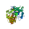
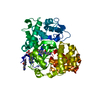
| Structure viewer | Molecule: MolmilJmol/JSmol |
|---|
- Downloads & links
-Download
| PDBx/mmCIF format | 7bj1.cif.gz | 142.4 KB | Display | PDBx/mmCIF format |
|---|---|---|---|---|
| PDB format | pdb7bj1.ent.gz | 86.4 KB | Display | PDB format |
| PDBx/mmJSON format | 7bj1.json.gz | Tree view | PDBx/mmJSON format | |
| Others |  Other downloads Other downloads |
-Validation report
| Arichive directory | https://data.pdbj.org/pub/pdb/validation_reports/bj/7bj1ftp://data.pdbj.org/pub/pdb/validation_reports/bj/7bj1 | HTTPS FTP |
|---|
-Related structure data
| Related structure data |  5cclS S: Starting model for refinement |
|---|---|
| Similar structure data |


-Links
 PDBj
PDBj

- Assembly
| Deposited unit | 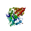
| ||||||||||||
|---|---|---|---|---|---|---|---|---|---|---|---|---|---|
| 1 |
| ||||||||||||
| Unit cell |
|
-Components
-Protein , 1 types, 1 molecules A
| #1: Protein | Mass: 49460.402 Da / Num. of mol.: 1 / Mutation: K13N, K140R Source method: isolated from a genetically manipulated source Source: (gene. exp.) Homo sapiens (human) / Gene: SMYD3, ZMYND1, ZNFN3A1 / Production host:  Escherichia coli BL21(DE3) (bacteria) Escherichia coli BL21(DE3) (bacteria)References: UniProt: Q9H7B4, [histone H3]-lysine4 N-trimethyltransferase |
|---|
-Non-polymers , 6 types, 532 molecules 


| #2: Chemical | ChemComp-SAM / S-Adenosyl methionine Mass: 398.437 Da / Num. of mol.: 1 / Source method: obtained synthetically / Formula: C15H22N6O5S Mass: 398.437 Da / Num. of mol.: 1 / Source method: obtained synthetically / Formula: C15H22N6O5S | ||||||
|---|---|---|---|---|---|---|---|
| #3: Chemical | ChemComp-QKT /  Mass: 397.467 Da / Num. of mol.: 1 / Source method: obtained synthetically / Formula: C22H27N3O4 / Feature type: SUBJECT OF INVESTIGATION Mass: 397.467 Da / Num. of mol.: 1 / Source method: obtained synthetically / Formula: C22H27N3O4 / Feature type: SUBJECT OF INVESTIGATION | ||||||
| #4: Chemical |  Mass: 65.409 Da / Num. of mol.: 3 / Source method: obtained synthetically / Formula: Zn Mass: 65.409 Da / Num. of mol.: 3 / Source method: obtained synthetically / Formula: Zn#5: Chemical | Acetate Mass: 59.044 Da / Num. of mol.: 2 / Source method: obtained synthetically / Formula: C2H3O2 Mass: 59.044 Da / Num. of mol.: 2 / Source method: obtained synthetically / Formula: C2H3O2#6: Chemical | Glycerol Mass: 92.094 Da / Num. of mol.: 2 / Source method: obtained synthetically / Formula: C3H8O3 Mass: 92.094 Da / Num. of mol.: 2 / Source method: obtained synthetically / Formula: C3H8O3#7: Water | ChemComp-HOH / | WaterMass: 18.015 Da / Num. of mol.: 523 / Source method: isolated from a natural source / Formula: H2O |
-Details
| Has ligand of interest | Y |
|---|
-Experimental details
-Experiment
| Experiment | Method: X-RAY DIFFRACTION / Number of used crystals: 1 |
|---|
- Sample preparation
| Crystal | Density Matthews: 2.18 Å3/Da / Density % sol: 43.62 % |
|---|---|
| Crystal grow | Temperature: 293 K / Method: vapor diffusion, hanging drop / pH: 8.25 Details: Protein: 50 mM Tris, 150 mM NaCl, 1 mM (S)-diperodon, 10% DMSO, pH 8.0; reservoir: 100 mM Tris, 50 mM magnesium acetate, 11% PEG3350, pH 8.25 |
-Data collection
| Diffraction | Mean temperature: 100 K / Serial crystal experiment: N |
|---|---|
| Diffraction source | Source: SYNCHROTRON / Site: ESRF  / Beamline: ID29 / Wavelength: 0.976 Å / Beamline: ID29 / Wavelength: 0.976 Å |
| Detector | Type: DECTRIS PILATUS 6M-F / Detector: PIXEL / Date: Jun 19, 2018 |
| Radiation | Protocol: SINGLE WAVELENGTH / Monochromatic (M) / Laue (L): M / Scattering type: x-ray |
| Radiation wavelength | Wavelength: 0.976 Å / Relative weight: 1 |
| Reflection | Resolution: 1.61→56.24 Å / Num. obs: 56743 / % possible obs: 99.9 % / Redundancy: 5.1 % / CC1/2: 0.999 / Rmerge(I) obs: 0.067 / Rpim(I) all: 0.033 / Rrim(I) all: 0.075 / Net I/σ(I): 12.3 |
| Reflection shell | Resolution: 1.61→1.67 Å / Redundancy: 4.8 % / Rmerge(I) obs: 1.528 / Num. unique obs: 5473 / CC1/2: 0.466 / Rpim(I) all: 0.765 / Rrim(I) all: 1.714 / % possible all: 99.8 |
- Processing
| Software |
| ||||||||||||||||||||||||||||||||||||||||||||||||||||||||||||||||||||||||||||||||||||||||||||||||||||||||||||||||||||||||||||||||||||||||||||
|---|---|---|---|---|---|---|---|---|---|---|---|---|---|---|---|---|---|---|---|---|---|---|---|---|---|---|---|---|---|---|---|---|---|---|---|---|---|---|---|---|---|---|---|---|---|---|---|---|---|---|---|---|---|---|---|---|---|---|---|---|---|---|---|---|---|---|---|---|---|---|---|---|---|---|---|---|---|---|---|---|---|---|---|---|---|---|---|---|---|---|---|---|---|---|---|---|---|---|---|---|---|---|---|---|---|---|---|---|---|---|---|---|---|---|---|---|---|---|---|---|---|---|---|---|---|---|---|---|---|---|---|---|---|---|---|---|---|---|---|---|---|
| Refinement | Method to determine structure: MOLECULAR REPLACEMENT Starting model: 5CCL Resolution: 1.61→44.78 Å / SU ML: 0.2463 / Cross valid method: FREE R-VALUE / σ(F): 1.35 / Phase error: 23.3432 Stereochemistry target values: GeoStd + Monomer Library + CDL v1.2
| ||||||||||||||||||||||||||||||||||||||||||||||||||||||||||||||||||||||||||||||||||||||||||||||||||||||||||||||||||||||||||||||||||||||||||||
| Solvent computation | Shrinkage radii: 0.9 Å / VDW probe radii: 1.11 Å / Solvent model: FLAT BULK SOLVENT MODEL | ||||||||||||||||||||||||||||||||||||||||||||||||||||||||||||||||||||||||||||||||||||||||||||||||||||||||||||||||||||||||||||||||||||||||||||
| Displacement parameters | Biso mean: 30.1 Å2 | ||||||||||||||||||||||||||||||||||||||||||||||||||||||||||||||||||||||||||||||||||||||||||||||||||||||||||||||||||||||||||||||||||||||||||||
| Refinement step | Cycle: LAST / Resolution: 1.61→44.78 Å
| ||||||||||||||||||||||||||||||||||||||||||||||||||||||||||||||||||||||||||||||||||||||||||||||||||||||||||||||||||||||||||||||||||||||||||||
| Refine LS restraints |
| ||||||||||||||||||||||||||||||||||||||||||||||||||||||||||||||||||||||||||||||||||||||||||||||||||||||||||||||||||||||||||||||||||||||||||||
| LS refinement shell |
|
