 Movie
Movie Controller

[English] 日本語
 Yorodumi
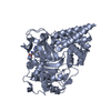
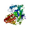
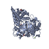
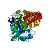
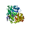
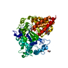
- PDB-6p6g: Co-crystal Structure of human SMYD3 with Isoxazole Amides Inhibitors -
+ Open data

- Basic information
| Entry | Database: PDB / ID: 6p6g | ||||||
|---|---|---|---|---|---|---|---|
| Title | Co-crystal Structure of human SMYD3 with Isoxazole Amides Inhibitors | ||||||
 Components Components | Histone-lysine N-methyltransferase SMYD3 | ||||||
 Keywords Keywords | TRANSFERASE/TRANSFERASE INHIBITOR /  methyltransferase / oncology / inhibitor / TRANSFERASE-TRANSFERASE INHIBITOR complex methyltransferase / oncology / inhibitor / TRANSFERASE-TRANSFERASE INHIBITOR complex | ||||||
| Function / homology |  Function and homology information Function and homology informationhistone H3K36 dimethyltransferase activity / histone H4 methyltransferase activity / [histone H3]-lysine4 N-trimethyltransferase / myotube cell development / histone H3K4 trimethyltransferase activity / RNA polymerase II intronic transcription regulatory region sequence-specific DNA binding / RNA polymerase II complex binding / cellular response to dexamethasone stimulus / establishment of protein localization / PKMTs methylate histone lysines ...histone H3K36 dimethyltransferase activity / histone H4 methyltransferase activity / [histone H3]-lysine4 N-trimethyltransferase / myotube cell development / histone H3K4 trimethyltransferase activity / RNA polymerase II intronic transcription regulatory region sequence-specific DNA binding / RNA polymerase II complex binding / cellular response to dexamethasone stimulus / establishment of protein localization / PKMTs methylate histone lysines / nucleosome assembly / positive regulation of peptidyl-serine phosphorylation / methylation / RNA polymerase II cis-regulatory region sequence-specific DNA binding / positive regulation of transcription by RNA polymerase II / nucleoplasm / metal ion binding / nucleus / cytosolSimilarity search - Function | ||||||
| Biological species |  Homo sapiens (human) Homo sapiens (human) | ||||||
| Method | X-RAY DIFFRACTION / SYNCHROTRON / MOLECULAR REPLACEMENT / Resolution: 1.59 Å | ||||||
 Authors Authors | Elkins, P.A. / Wang, L. | ||||||
 Citation Citation | Journal: Acs Med.Chem.Lett. / Year: 2020 Title: Discovery of Isoxazole Amides as Potent and Selective SMYD3 Inhibitors. Authors: Su, D.S. / Qu, J. / Schulz, M. / Blackledge, C.W. / Yu, H. / Zeng, J. / Burgess, J. / Reif, A. / Stern, M. / Nagarajan, R. / Pappalardi, M.B. / Wong, K. / Graves, A.P. / Bonnette, W. / Wang, ...Authors: Su, D.S. / Qu, J. / Schulz, M. / Blackledge, C.W. / Yu, H. / Zeng, J. / Burgess, J. / Reif, A. / Stern, M. / Nagarajan, R. / Pappalardi, M.B. / Wong, K. / Graves, A.P. / Bonnette, W. / Wang, L. / Elkins, P. / Knapp-Reed, B. / Carson, J.D. / McHugh, C. / Mohammad, H. / Kruger, R. / Luengo, J. / Heerding, D.A. / Creasy, C.L. | ||||||
| History |
|
- Structure visualization
| Structure viewer | Molecule: MolmilJmol/JSmol |
|---|
- Downloads & links
-Download
| PDBx/mmCIF format | 6p6g.cif.gz | 114.3 KB | Display | PDBx/mmCIF format |
|---|---|---|---|---|
| PDB format | pdb6p6g.ent.gz | 83 KB | Display | PDB format |
| PDBx/mmJSON format | 6p6g.json.gz | Tree view | PDBx/mmJSON format | |
| Others |  Other downloads Other downloads |
-Validation report
| Arichive directory | https://data.pdbj.org/pub/pdb/validation_reports/p6/6p6gftp://data.pdbj.org/pub/pdb/validation_reports/p6/6p6g | HTTPS FTP |
|---|
-Related structure data


-Links
 PDBj
PDBj
- Assembly
| Deposited unit | 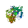
| ||||||||
|---|---|---|---|---|---|---|---|---|---|
| 1 |
| ||||||||
| Unit cell |
|
-Components
-Protein , 1 types, 1 molecules A
| #1: Protein | Mass: 49542.520 Da / Num. of mol.: 1 Source method: isolated from a genetically manipulated source Source: (gene. exp.) Homo sapiens (human) / Gene: SMYD3, ZMYND1, ZNFN3A1 / Cell line (production host): Sf9 / Production host:  unidentified baculovirus unidentified baculovirusReferences: UniProt: Q9H7B4, histone-lysine N-methyltransferase |
|---|
-Non-polymers , 6 types, 401 molecules 


| #2: Chemical |  Mass: 65.409 Da / Num. of mol.: 3 / Source method: obtained synthetically / Formula: Zn Mass: 65.409 Da / Num. of mol.: 3 / Source method: obtained synthetically / Formula: Zn#3: Chemical |  Mass: 24.305 Da / Num. of mol.: 2 / Source method: obtained synthetically / Formula: Mg Mass: 24.305 Da / Num. of mol.: 2 / Source method: obtained synthetically / Formula: Mg#4: Chemical | Glycerol Mass: 92.094 Da / Num. of mol.: 3 / Source method: obtained synthetically / Formula: C3H8O3 Mass: 92.094 Da / Num. of mol.: 3 / Source method: obtained synthetically / Formula: C3H8O3#5: Chemical | ChemComp-SAH / | S-Adenosyl-L-homocysteine Type: L-peptide linking / Mass: 384.411 Da / Num. of mol.: 1 / Source method: obtained synthetically / Formula: C14H20N6O5S Type: L-peptide linking / Mass: 384.411 Da / Num. of mol.: 1 / Source method: obtained synthetically / Formula: C14H20N6O5S#6: Chemical | ChemComp-LUP / |  Mass: 520.609 Da / Num. of mol.: 1 / Source method: obtained synthetically / Formula: C23H35F3N4O4S / Feature type: SUBJECT OF INVESTIGATION Mass: 520.609 Da / Num. of mol.: 1 / Source method: obtained synthetically / Formula: C23H35F3N4O4S / Feature type: SUBJECT OF INVESTIGATION#7: Water | ChemComp-HOH / | WaterMass: 18.015 Da / Num. of mol.: 391 / Source method: isolated from a natural source / Formula: H2O |
|---|
-Experimental details
-Experiment
| Experiment | Method: X-RAY DIFFRACTION / Number of used crystals: 1 |
|---|
- Sample preparation
| Crystal | Density Matthews: 2.22 Å3/Da / Density % sol: 44.53 % |
|---|---|
| Crystal grow | Temperature: 295 K / Method: vapor diffusion, hanging drop Details: Sitting Drop, Linbro tray, room temp, reservoir is 10% PEG 3350, 0.2M MgOAc , protein:reservoir is 1:1, streak seed soaking: 2ul 20%PEG 3350, 0.4M MgOAc added to 2ul protein, mix and add 0. ...Details: Sitting Drop, Linbro tray, room temp, reservoir is 10% PEG 3350, 0.2M MgOAc , protein:reservoir is 1:1, streak seed soaking: 2ul 20%PEG 3350, 0.4M MgOAc added to 2ul protein, mix and add 0.3ul to drop, store at 295 |
-Data collection
| Diffraction | Mean temperature: 100 K / Serial crystal experiment: N | ||||||||||||||||||||||||
|---|---|---|---|---|---|---|---|---|---|---|---|---|---|---|---|---|---|---|---|---|---|---|---|---|---|
| Diffraction source | Source: SYNCHROTRON / Site: APS  / Beamline: 21-ID-D / Wavelength: 1.07805 Å / Beamline: 21-ID-D / Wavelength: 1.07805 Å | ||||||||||||||||||||||||
| Detector | Type: MAR scanner 345 mm plate / Detector: IMAGE PLATE / Date: Aug 5, 2015 | ||||||||||||||||||||||||
| Radiation | Protocol: SINGLE WAVELENGTH / Monochromatic (M) / Laue (L): M / Scattering type: x-ray | ||||||||||||||||||||||||
| Radiation wavelength | Wavelength: 1.07805 Å / Relative weight: 1 | ||||||||||||||||||||||||
| Reflection | Resolution: 1.59→66.64 Å / Num. obs: 60184 / % possible obs: 99.6 % / Redundancy: 6.9 % / CC1/2: 0.999 / Rmerge(I) obs: 0.071 / Rpim(I) all: 0.029 / Rrim(I) all: 0.077 / Net I/σ(I): 15 / Num. measured all: 413998 | ||||||||||||||||||||||||
| Reflection shell | Diffraction-ID: 1
|
- Processing
| Software |
| |||||||||||||||||||||||||||||||||||||||||||||||||||||||||||||||||||||||||||||||||||||||||||||||||||||||||||||||||||||||||||||||||||||||||||||||||||||||||||||||||||||||||||||||||||||||||||||||||||||||||||||||||||||||||
|---|---|---|---|---|---|---|---|---|---|---|---|---|---|---|---|---|---|---|---|---|---|---|---|---|---|---|---|---|---|---|---|---|---|---|---|---|---|---|---|---|---|---|---|---|---|---|---|---|---|---|---|---|---|---|---|---|---|---|---|---|---|---|---|---|---|---|---|---|---|---|---|---|---|---|---|---|---|---|---|---|---|---|---|---|---|---|---|---|---|---|---|---|---|---|---|---|---|---|---|---|---|---|---|---|---|---|---|---|---|---|---|---|---|---|---|---|---|---|---|---|---|---|---|---|---|---|---|---|---|---|---|---|---|---|---|---|---|---|---|---|---|---|---|---|---|---|---|---|---|---|---|---|---|---|---|---|---|---|---|---|---|---|---|---|---|---|---|---|---|---|---|---|---|---|---|---|---|---|---|---|---|---|---|---|---|---|---|---|---|---|---|---|---|---|---|---|---|---|---|---|---|---|---|---|---|---|---|---|---|---|---|---|---|---|---|---|---|---|
| Refinement | Method to determine structure: MOLECULAR REPLACEMENT / Resolution: 1.59→56.614 Å / SU ML: 0.19 / Cross valid method: THROUGHOUT / σ(F): 1.36 / Phase error: 20.9
| |||||||||||||||||||||||||||||||||||||||||||||||||||||||||||||||||||||||||||||||||||||||||||||||||||||||||||||||||||||||||||||||||||||||||||||||||||||||||||||||||||||||||||||||||||||||||||||||||||||||||||||||||||||||||
| Solvent computation | Shrinkage radii: 0.9 Å / VDW probe radii: 1.11 Å | |||||||||||||||||||||||||||||||||||||||||||||||||||||||||||||||||||||||||||||||||||||||||||||||||||||||||||||||||||||||||||||||||||||||||||||||||||||||||||||||||||||||||||||||||||||||||||||||||||||||||||||||||||||||||
| Displacement parameters | Biso max: 89.92 Å2 / Biso mean: 23.7103 Å2 / Biso min: 9.08 Å2 | |||||||||||||||||||||||||||||||||||||||||||||||||||||||||||||||||||||||||||||||||||||||||||||||||||||||||||||||||||||||||||||||||||||||||||||||||||||||||||||||||||||||||||||||||||||||||||||||||||||||||||||||||||||||||
| Refinement step | Cycle: final / Resolution: 1.59→56.614 Å
| |||||||||||||||||||||||||||||||||||||||||||||||||||||||||||||||||||||||||||||||||||||||||||||||||||||||||||||||||||||||||||||||||||||||||||||||||||||||||||||||||||||||||||||||||||||||||||||||||||||||||||||||||||||||||
| LS refinement shell | Refine-ID: X-RAY DIFFRACTION / Rfactor Rfree error: 0 / Total num. of bins used: 30
|
