 Movie
Movie Controller


+ Open data

- Basic information
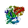
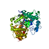
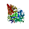
| Entry | Database: PDB / ID: 6ijl | ||||||
|---|---|---|---|---|---|---|---|
| Title | Crystal structure of SmyD3 in complex with covalent inhibitor 5 | ||||||
 Components Components | Histone-lysine N-methyltransferase SMYD3 | ||||||
 Keywords Keywords | TRANSFERASE/INHIBITOR / covalent inhibitor /  methyltransferase / methyltransferase inhibitor / TRANSFERASE-INHIBITOR complex methyltransferase / methyltransferase inhibitor / TRANSFERASE-INHIBITOR complex | ||||||
| Function / homology |  Function and homology information Function and homology informationhistone H3K36 dimethyltransferase activity / histone H4 methyltransferase activity / [histone H3]-lysine4 N-trimethyltransferase / myotube cell development / histone H3K4 trimethyltransferase activity / RNA polymerase II intronic transcription regulatory region sequence-specific DNA binding / RNA polymerase II complex binding / cellular response to dexamethasone stimulus / establishment of protein localization / PKMTs methylate histone lysines ...histone H3K36 dimethyltransferase activity / histone H4 methyltransferase activity / [histone H3]-lysine4 N-trimethyltransferase / myotube cell development / histone H3K4 trimethyltransferase activity / RNA polymerase II intronic transcription regulatory region sequence-specific DNA binding / RNA polymerase II complex binding / cellular response to dexamethasone stimulus / establishment of protein localization / PKMTs methylate histone lysines / nucleosome assembly / positive regulation of peptidyl-serine phosphorylation / methylation / RNA polymerase II cis-regulatory region sequence-specific DNA binding / positive regulation of transcription by RNA polymerase II / nucleoplasm / metal ion binding / nucleus / cytosolSimilarity search - Function | ||||||
| Biological species |  Homo sapiens (human) Homo sapiens (human) | ||||||
| Method | X-RAY DIFFRACTION / MOLECULAR REPLACEMENT / Resolution: 2.351 Å | ||||||
 Authors Authors | Baburajendran, N. / Joy, J. | ||||||
 Citation Citation | Journal: Acs Med.Chem.Lett. / Year: 2019 Title: Discovery of Irreversible Inhibitors Targeting Histone Methyltransferase, SMYD3. Authors: Huang, C. / Liew, S.S. / Lin, G.R. / Poulsen, A. / Ang, M.J.Y. / Chia, B.C.S. / Chew, S.Y. / Kwek, Z.P. / Wee, J.L.K. / Ong, E.H. / Retna, P. / Baburajendran, N. / Li, R. / Yu, W. / Koh- ...Authors: Huang, C. / Liew, S.S. / Lin, G.R. / Poulsen, A. / Ang, M.J.Y. / Chia, B.C.S. / Chew, S.Y. / Kwek, Z.P. / Wee, J.L.K. / Ong, E.H. / Retna, P. / Baburajendran, N. / Li, R. / Yu, W. / Koh-Stenta, X. / Ngo, A. / Manesh, S. / Fulwood, J. / Ke, Z. / Chung, H.H. / Sepramaniam, S. / Chew, X.H. / Dinie, N. / Lee, M.A. / Chew, Y.S. / Low, C.B. / Pendharkar, V. / Manoharan, V. / Vuddagiri, S. / Sangthongpitag, K. / Joy, J. / Matter, A. / Hill, J. / Keller, T.H. / Foo, K. | ||||||
| History |
|
- Structure visualization
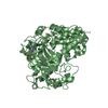
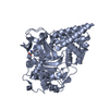
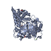
| Structure viewer | Molecule: MolmilJmol/JSmol |
|---|
- Downloads & links
-Download
| PDBx/mmCIF format | 6ijl.cif.gz | 186.1 KB | Display | PDBx/mmCIF format |
|---|---|---|---|---|
| PDB format | pdb6ijl.ent.gz | 144.9 KB | Display | PDB format |
| PDBx/mmJSON format | 6ijl.json.gz | Tree view | PDBx/mmJSON format | |
| Others |  Other downloads Other downloads |
-Validation report
| Arichive directory | https://data.pdbj.org/pub/pdb/validation_reports/ij/6ijlftp://data.pdbj.org/pub/pdb/validation_reports/ij/6ijl | HTTPS FTP |
|---|
-Related structure data


-Links
 PDBj
PDBj

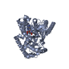
- Assembly
| Deposited unit | 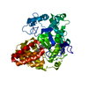
| ||||||||
|---|---|---|---|---|---|---|---|---|---|
| 1 |
| ||||||||
| Unit cell |
|
-Components
| #1: Protein | Mass: 49178.121 Da / Num. of mol.: 1 / Mutation: K13N, K140R Source method: isolated from a genetically manipulated source Source: (gene. exp.) Homo sapiens (human) / Gene: SMYD3 / Production host:  Escherichia coli (E. coli) Escherichia coli (E. coli)References: UniProt: Q9H7B4, histone-lysine N-methyltransferase | ||||||
|---|---|---|---|---|---|---|---|
| #2: Chemical | ChemComp-A8U /   Mass: 472.579 Da / Num. of mol.: 1 / Source method: obtained synthetically / Formula: C28H32N4O3 / Feature type: SUBJECT OF INVESTIGATION Mass: 472.579 Da / Num. of mol.: 1 / Source method: obtained synthetically / Formula: C28H32N4O3 / Feature type: SUBJECT OF INVESTIGATION | ||||||
| #3: Chemical |   Mass: 65.409 Da / Num. of mol.: 3 / Source method: obtained synthetically / Formula: Zn Mass: 65.409 Da / Num. of mol.: 3 / Source method: obtained synthetically / Formula: Zn#4: Chemical | ChemComp-SAM / | S-Adenosyl methionine  Mass: 398.437 Da / Num. of mol.: 1 / Source method: obtained synthetically / Formula: C15H22N6O5S Mass: 398.437 Da / Num. of mol.: 1 / Source method: obtained synthetically / Formula: C15H22N6O5S#5: Water | ChemComp-HOH / | Water Mass: 18.015 Da / Num. of mol.: 100 / Source method: isolated from a natural source / Formula: H2O Mass: 18.015 Da / Num. of mol.: 100 / Source method: isolated from a natural source / Formula: H2OHas ligand of interest | Y | |
-Experimental details
-Experiment
| Experiment | Method: X-RAY DIFFRACTION / Number of used crystals: 1 |
|---|
- Sample preparation
| Crystal | Density Matthews: 2.24 Å3/Da / Density % sol: 45.08 % |
|---|---|
| Crystal grow | Temperature: 293 K / Method: vapor diffusion, hanging drop / Details: 0.2M Magnesium acetate, 17% PEG 3350 |
-Data collection
| Diffraction | Mean temperature: 100 K / Serial crystal experiment: N | |||||||||||||||||||||||||||||||||||||||||||||||||||||||||||||||||||||||||||||||||||||||||||||||||||||||||||||||||||||||||||||||||||||||||||||||||||||||||||||||||||||||||||||||||||||||||||||
|---|---|---|---|---|---|---|---|---|---|---|---|---|---|---|---|---|---|---|---|---|---|---|---|---|---|---|---|---|---|---|---|---|---|---|---|---|---|---|---|---|---|---|---|---|---|---|---|---|---|---|---|---|---|---|---|---|---|---|---|---|---|---|---|---|---|---|---|---|---|---|---|---|---|---|---|---|---|---|---|---|---|---|---|---|---|---|---|---|---|---|---|---|---|---|---|---|---|---|---|---|---|---|---|---|---|---|---|---|---|---|---|---|---|---|---|---|---|---|---|---|---|---|---|---|---|---|---|---|---|---|---|---|---|---|---|---|---|---|---|---|---|---|---|---|---|---|---|---|---|---|---|---|---|---|---|---|---|---|---|---|---|---|---|---|---|---|---|---|---|---|---|---|---|---|---|---|---|---|---|---|---|---|---|---|---|---|---|---|---|---|
| Diffraction source | Source: ROTATING ANODE / Type: RIGAKU MICROMAX-007 / Wavelength: 1.5418 Å | |||||||||||||||||||||||||||||||||||||||||||||||||||||||||||||||||||||||||||||||||||||||||||||||||||||||||||||||||||||||||||||||||||||||||||||||||||||||||||||||||||||||||||||||||||||||||||||
| Detector | Type: RIGAKU SATURN 944+ / Detector: CCD / Date: Apr 3, 2018 | |||||||||||||||||||||||||||||||||||||||||||||||||||||||||||||||||||||||||||||||||||||||||||||||||||||||||||||||||||||||||||||||||||||||||||||||||||||||||||||||||||||||||||||||||||||||||||||
| Radiation | Protocol: SINGLE WAVELENGTH / Monochromatic (M) / Laue (L): M / Scattering type: x-ray | |||||||||||||||||||||||||||||||||||||||||||||||||||||||||||||||||||||||||||||||||||||||||||||||||||||||||||||||||||||||||||||||||||||||||||||||||||||||||||||||||||||||||||||||||||||||||||||
| Radiation wavelength | Wavelength: 1.5418 Å / Relative weight: 1 | |||||||||||||||||||||||||||||||||||||||||||||||||||||||||||||||||||||||||||||||||||||||||||||||||||||||||||||||||||||||||||||||||||||||||||||||||||||||||||||||||||||||||||||||||||||||||||||
| Reflection | Resolution: 2.35→50 Å / Num. obs: 18706 / % possible obs: 99.3 % / Redundancy: 6.9 % / Biso Wilson estimate: 25.51 Å2 / Rmerge(I) obs: 0.136 / Rpim(I) all: 0.055 / Rrim(I) all: 0.147 / Χ2: 0.973 / Net I/σ(I): 8.1 / Num. measured all: 128300 | |||||||||||||||||||||||||||||||||||||||||||||||||||||||||||||||||||||||||||||||||||||||||||||||||||||||||||||||||||||||||||||||||||||||||||||||||||||||||||||||||||||||||||||||||||||||||||||
| Reflection shell | Diffraction-ID: 1
|
- Processing
| Software |
| ||||||||||||||||||||||||||||||||||||||||||||||||||||||||||||||||||||||||||||||||||||
|---|---|---|---|---|---|---|---|---|---|---|---|---|---|---|---|---|---|---|---|---|---|---|---|---|---|---|---|---|---|---|---|---|---|---|---|---|---|---|---|---|---|---|---|---|---|---|---|---|---|---|---|---|---|---|---|---|---|---|---|---|---|---|---|---|---|---|---|---|---|---|---|---|---|---|---|---|---|---|---|---|---|---|---|---|---|
| Refinement | Method to determine structure: MOLECULAR REPLACEMENT / Resolution: 2.351→41.712 Å / SU ML: 0.27 / Cross valid method: THROUGHOUT / σ(F): 1.33 / Phase error: 30.15 / Stereochemistry target values: ML
| ||||||||||||||||||||||||||||||||||||||||||||||||||||||||||||||||||||||||||||||||||||
| Solvent computation | Shrinkage radii: 0.9 Å / VDW probe radii: 1.11 Å / Solvent model: FLAT BULK SOLVENT MODEL | ||||||||||||||||||||||||||||||||||||||||||||||||||||||||||||||||||||||||||||||||||||
| Displacement parameters | Biso max: 84.25 Å2 / Biso mean: 27.8926 Å2 / Biso min: 11.72 Å2 | ||||||||||||||||||||||||||||||||||||||||||||||||||||||||||||||||||||||||||||||||||||
| Refinement step | Cycle: final / Resolution: 2.351→41.712 Å
| ||||||||||||||||||||||||||||||||||||||||||||||||||||||||||||||||||||||||||||||||||||
| Refine LS restraints |
| ||||||||||||||||||||||||||||||||||||||||||||||||||||||||||||||||||||||||||||||||||||
| LS refinement shell | Refine-ID: X-RAY DIFFRACTION / Rfactor Rfree error: 0
| ||||||||||||||||||||||||||||||||||||||||||||||||||||||||||||||||||||||||||||||||||||
| Refinement TLS params. | Method: refined / Origin x: 73.5347 Å / Origin y: 131.9699 Å / Origin z: 120.3688 Å
| ||||||||||||||||||||||||||||||||||||||||||||||||||||||||||||||||||||||||||||||||||||
| Refinement TLS group |
|
