 Movie
Movie Controller

[English] 日本語
 Yorodumi
- PDB-6zsq: Crystal structure of the Cisplatin beta-Lactoglobulin adduct form... -
+ Open data

- Basic information
| Entry | Database: PDB / ID: 6zsq | ||||||
|---|---|---|---|---|---|---|---|
| Title | Crystal structure of the Cisplatin beta-Lactoglobulin adduct formed after 18 h of soaking | ||||||
 Components Components | Beta-lactoglobulin | ||||||
 Keywords Keywords | TRANSPORT PROTEIN / Complex / Drug delivery system / Cisplatin | ||||||
| Function / homology |  Function and homology informationretinol binding / long-chain fatty acid binding / extracellular space / identical protein binding Function and homology informationretinol binding / long-chain fatty acid binding / extracellular space / identical protein bindingSimilarity search - Function | ||||||
| Biological species |  Bos taurus (cattle) Bos taurus (cattle) | ||||||
| Method | X-RAY DIFFRACTION / MOLECULAR REPLACEMENT / Resolution: 2.004 Å | ||||||
 Authors Authors | Balasco, N. / Ferraro, G. / Merlino, A. | ||||||
 Citation Citation | Journal: Dalton Trans / Year: 2020 Title: Cisplatin binding to beta-lactoglobulin: a structural study. Authors: Balasco, N. / Ferraro, G. / Loreto, D. / Iacobucci, I. / Monti, M. / Merlino, A. | ||||||
| History |
|
- Structure visualization
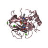
| Structure viewer | Molecule: MolmilJmol/JSmol |
|---|
- Downloads & links
-Download
| PDBx/mmCIF format | 6zsq.cif.gz | 84 KB | Display | PDBx/mmCIF format |
|---|---|---|---|---|
| PDB format | pdb6zsq.ent.gz | Display | PDB format | |
| PDBx/mmJSON format | 6zsq.json.gz | Tree view | PDBx/mmJSON format | |
| Others |  Other downloads Other downloads |
-Validation report
| Arichive directory | https://data.pdbj.org/pub/pdb/validation_reports/zs/6zsqftp://data.pdbj.org/pub/pdb/validation_reports/zs/6zsq | HTTPS FTP |
|---|
-Related structure data
| Related structure data | 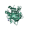 6zsrC  1bebS S: Starting model for refinement C: citing same article ( |
|---|---|
| Similar structure data |


-Links
 PDBj
PDBj


- Assembly
| Deposited unit | 
| ||||||||
|---|---|---|---|---|---|---|---|---|---|
| 1 | 
| ||||||||
| 2 | 
| ||||||||
| Unit cell |
|
-Components
| #1: Protein | / Beta-LG Mass: 18329.229 Da / Num. of mol.: 2 / Source method: isolated from a natural source / Source: (natural) Bos taurus (cattle) / References: UniProt: P02754#2: Chemical | ChemComp-NH3 / | Ammonia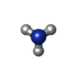  Mass: 17.031 Da / Num. of mol.: 1 / Source method: obtained synthetically / Formula: NH3 / Feature type: SUBJECT OF INVESTIGATION Mass: 17.031 Da / Num. of mol.: 1 / Source method: obtained synthetically / Formula: NH3 / Feature type: SUBJECT OF INVESTIGATION#3: Chemical |   Mass: 195.078 Da / Num. of mol.: 3 / Source method: obtained synthetically / Formula: Pt / Feature type: SUBJECT OF INVESTIGATION Mass: 195.078 Da / Num. of mol.: 3 / Source method: obtained synthetically / Formula: Pt / Feature type: SUBJECT OF INVESTIGATION#4: Chemical | Sulfate  Mass: 96.063 Da / Num. of mol.: 2 / Source method: obtained synthetically / Formula: SO4 Mass: 96.063 Da / Num. of mol.: 2 / Source method: obtained synthetically / Formula: SO4#5: Water | ChemComp-HOH / | Water Mass: 18.015 Da / Num. of mol.: 165 / Source method: isolated from a natural source / Formula: H2O Mass: 18.015 Da / Num. of mol.: 165 / Source method: isolated from a natural source / Formula: H2OHas ligand of interest | Y | |
|---|
-Experimental details
-Experiment
| Experiment | Method: X-RAY DIFFRACTION / Number of used crystals: 1 |
|---|
- Sample preparation
| Crystal | Density Matthews: 2.16 Å3/Da / Density % sol: 43.12 % |
|---|---|
| Crystal grow | Temperature: 293 K / Method: vapor diffusion, sitting drop / pH: 6.9 Details: Crystals of beta-Lactoglobulin were grown using 3.0 M (NH4)2SO4, 0.1 M sodium/potassium phosphate buffer pH 6.9 as reservoir. Protein crystallizes at a concentration of 18 mg/mL. Crystals of ...Details: Crystals of beta-Lactoglobulin were grown using 3.0 M (NH4)2SO4, 0.1 M sodium/potassium phosphate buffer pH 6.9 as reservoir. Protein crystallizes at a concentration of 18 mg/mL. Crystals of beta-Lactoglobulin were soaked in a solution consisting of 3.0 M (NH4)2SO4, 0.1 M sodium/potassium phosphate buffer (pH 6.9) with 5 mM CDDP (about 1:3 protein to metal ratio). |
-Data collection
| Diffraction | Mean temperature: 100 K / Serial crystal experiment: N |
|---|---|
| Diffraction source | Source: ROTATING ANODE / Type: RIGAKU MICROMAX-007 / Wavelength: 1.54 Å |
| Detector | Type: RIGAKU SATURN 944 / Detector: CCD / Date: Mar 16, 2018 |
| Radiation | Protocol: SINGLE WAVELENGTH / Monochromatic (M) / Laue (L): M / Scattering type: x-ray |
| Radiation wavelength | Wavelength: 1.54 Å / Relative weight: 1 |
| Reflection | Resolution: 2→23.23 Å / Num. obs: 17918 / % possible obs: 86.9 % / Redundancy: 2.3 % / CC1/2: 0.958 / Rmerge(I) obs: 0.06 / Net I/σ(I): 8.8 |
| Reflection shell | Resolution: 2→2.06 Å / Redundancy: 1.9 % / Rmerge(I) obs: 0.152 / Mean I/σ(I) obs: 3.9 / Num. unique obs: 2486 / CC1/2: 0.939 / % possible all: 92.4 |
- Processing
| Software |
| ||||||||||||||||||||||||||||||||||||||||||||||||||||||||||||||||||||||||||||||||||||||||||||||||||||||||||||||||||||||||||||||||||||||||||||||||||||||||||||||||
|---|---|---|---|---|---|---|---|---|---|---|---|---|---|---|---|---|---|---|---|---|---|---|---|---|---|---|---|---|---|---|---|---|---|---|---|---|---|---|---|---|---|---|---|---|---|---|---|---|---|---|---|---|---|---|---|---|---|---|---|---|---|---|---|---|---|---|---|---|---|---|---|---|---|---|---|---|---|---|---|---|---|---|---|---|---|---|---|---|---|---|---|---|---|---|---|---|---|---|---|---|---|---|---|---|---|---|---|---|---|---|---|---|---|---|---|---|---|---|---|---|---|---|---|---|---|---|---|---|---|---|---|---|---|---|---|---|---|---|---|---|---|---|---|---|---|---|---|---|---|---|---|---|---|---|---|---|---|---|---|---|---|
| Refinement | Method to determine structure: MOLECULAR REPLACEMENT Starting model: 1BEB Resolution: 2.004→23.228 Å / Cor.coef. Fo:Fc: 0.953 / Cor.coef. Fo:Fc free: 0.932 / SU B: 5.195 / SU ML: 0.141 / Cross valid method: FREE R-VALUE / ESU R: 0.265 / ESU R Free: 0.193 Details: Hydrogens have been added in their riding positions
| ||||||||||||||||||||||||||||||||||||||||||||||||||||||||||||||||||||||||||||||||||||||||||||||||||||||||||||||||||||||||||||||||||||||||||||||||||||||||||||||||
| Solvent computation | Ion probe radii: 0.8 Å / Shrinkage radii: 0.8 Å / VDW probe radii: 1.2 Å / Solvent model: MASK BULK SOLVENT | ||||||||||||||||||||||||||||||||||||||||||||||||||||||||||||||||||||||||||||||||||||||||||||||||||||||||||||||||||||||||||||||||||||||||||||||||||||||||||||||||
| Displacement parameters | Biso mean: 22.264 Å2
| ||||||||||||||||||||||||||||||||||||||||||||||||||||||||||||||||||||||||||||||||||||||||||||||||||||||||||||||||||||||||||||||||||||||||||||||||||||||||||||||||
| Refinement step | Cycle: LAST / Resolution: 2.004→23.228 Å
| ||||||||||||||||||||||||||||||||||||||||||||||||||||||||||||||||||||||||||||||||||||||||||||||||||||||||||||||||||||||||||||||||||||||||||||||||||||||||||||||||
| Refine LS restraints |
| ||||||||||||||||||||||||||||||||||||||||||||||||||||||||||||||||||||||||||||||||||||||||||||||||||||||||||||||||||||||||||||||||||||||||||||||||||||||||||||||||
| LS refinement shell |
|
