 Movie
Movie Controller


+ Open data

- Basic information
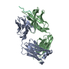
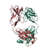
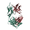
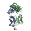
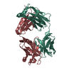
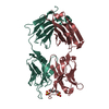
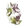
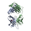
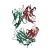
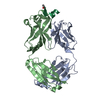
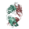
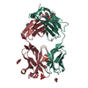
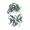
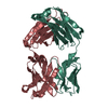
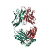
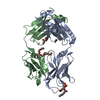
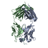
| Entry | Database: PDB / ID: 6yxk | |||||||||
|---|---|---|---|---|---|---|---|---|---|---|
| Title | Crystal structure of ACPA 3F3 in complex with cit-vimentin 59-74 | |||||||||
 Components Components |
| |||||||||
 Keywords Keywords |  IMMUNE SYSTEM / anti-citrullinated protein antibody Fab fragment / citrullinated vimentin IMMUNE SYSTEM / anti-citrullinated protein antibody Fab fragment / citrullinated vimentin | |||||||||
| Function / homology |  Function and homology information Function and homology informationkeratin filament binding / lens fiber cell development / intermediate filament organization / cellular response to muramyl dipeptide / structural constituent of eye lens / astrocyte development / Striated Muscle Contraction / RHOBTB1 GTPase cycle / intermediate filament / microtubule organizing center ...keratin filament binding / lens fiber cell development / intermediate filament organization / cellular response to muramyl dipeptide / structural constituent of eye lens / astrocyte development / Striated Muscle Contraction / RHOBTB1 GTPase cycle / intermediate filament / microtubule organizing center / intermediate filament cytoskeleton / cell leading edge / Bergmann glial cell differentiation / positive regulation of collagen biosynthetic process / Caspase-mediated cleavage of cytoskeletal proteins / phagocytic vesicle / regulation of mRNA stability / Late endosomal microautophagy / structural constituent of cytoskeleton / nuclear matrix / Aggrephagy / cellular response to type II interferon / Chaperone Mediated Autophagy / peroxisome / neuron projection development / double-stranded RNA binding / negative regulation of neuron projection development / scaffold protein binding / Interleukin-4 and Interleukin-13 signaling / cellular response to lipopolysaccharide / molecular adaptor activity / cytoskeleton / axon / protein domain specific binding / focal adhesion / positive regulation of gene expression / extracellular exosome / identical protein binding / plasma membrane / cytosol / cytoplasmSimilarity search - Function | |||||||||
| Biological species |  Homo sapiens (human) Homo sapiens (human) | |||||||||
| Method | X-RAY DIFFRACTION / SYNCHROTRON / MOLECULAR REPLACEMENT / Resolution: 2 Å | |||||||||
 Authors Authors | Ge, C. / Holmdahl, R. | |||||||||
| Funding support |  Sweden, 2items Sweden, 2items
| |||||||||
 Citation Citation | Journal: Sci Adv / Year: 2022 Title: Surface Ig variable domain glycosylation affects autoantigen binding and acts as threshold for human autoreactive B cell activation. Authors: Kissel, T. / Ge, C. / Hafkenscheid, L. / Kwekkeboom, J.C. / Slot, L.M. / Cavallari, M. / He, Y. / van Schie, K.A. / Vergroesen, R.D. / Kampstra, A.S.B. / Reijm, S. / Stoeken-Rijsbergen, G. / ...Authors: Kissel, T. / Ge, C. / Hafkenscheid, L. / Kwekkeboom, J.C. / Slot, L.M. / Cavallari, M. / He, Y. / van Schie, K.A. / Vergroesen, R.D. / Kampstra, A.S.B. / Reijm, S. / Stoeken-Rijsbergen, G. / Koeleman, C. / Voortman, L.M. / Heitman, L.H. / Xu, B. / Pruijn, G.J.M. / Wuhrer, M. / Rispens, T. / Huizinga, T.W.J. / Scherer, H.U. / Reth, M. / Holmdahl, R. / Toes, R.E.M. | |||||||||
| History |
|
- Structure visualization
| Structure viewer | Molecule: MolmilJmol/JSmol |
|---|
- Downloads & links
-Download
| PDBx/mmCIF format | 6yxk.cif.gz | 315.2 KB | Display | PDBx/mmCIF format |
|---|---|---|---|---|
| PDB format | pdb6yxk.ent.gz | 252.2 KB | Display | PDB format |
| PDBx/mmJSON format | 6yxk.json.gz | Tree view | PDBx/mmJSON format | |
| Others |  Other downloads Other downloads |
-Validation report
| Arichive directory | https://data.pdbj.org/pub/pdb/validation_reports/yx/6yxkftp://data.pdbj.org/pub/pdb/validation_reports/yx/6yxk | HTTPS FTP |
|---|
-Related structure data
| Related structure data |  6yxlC  6yxmC  5ocxS S: Starting model for refinement C: citing same article ( |
|---|---|
| Similar structure data |


-Links
 PDBj
PDBj


- Assembly
| Deposited unit | 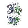
| ||||||||
|---|---|---|---|---|---|---|---|---|---|
| 1 |
| ||||||||
| Unit cell |
|
-Components
| #1: Antibody | Mass: 23772.783 Da / Num. of mol.: 1 Source method: isolated from a genetically manipulated source Source: (gene. exp.) Homo sapiens (human) / Plasmid: pCEP4 / Cell line (production host): Expi 293F(TM) / Production host: Homo sapiens (human) | ||||
|---|---|---|---|---|---|
| #2: Antibody | Mass: 24120.625 Da / Num. of mol.: 1 Source method: isolated from a genetically manipulated source Source: (gene. exp.) Homo sapiens (human) / Plasmid: pCEP4 / Cell line (production host): Expi 293F(TM) / Production host: Homo sapiens (human) | ||||
| #3: Protein/peptide | Mass: 1711.897 Da / Num. of mol.: 1 / Source method: obtained synthetically / Source: (synth.) Homo sapiens (human) / References: UniProt: P08670 | ||||
| #4: Sugar | N-Acetylglucosamine  Type: D-saccharide, beta linking / Mass: 221.208 Da / Num. of mol.: 3 Type: D-saccharide, beta linking / Mass: 221.208 Da / Num. of mol.: 3Source method: isolated from a genetically manipulated source Formula: C8H15NO6 #5: Water | ChemComp-HOH / | Water Mass: 18.015 Da / Num. of mol.: 303 / Source method: isolated from a natural source / Formula: H2O Mass: 18.015 Da / Num. of mol.: 303 / Source method: isolated from a natural source / Formula: H2OHas ligand of interest | Y | |
-Experimental details
-Experiment
| Experiment | Method: X-RAY DIFFRACTION / Number of used crystals: 1 |
|---|
- Sample preparation
| Crystal | Density Matthews: 3.03 Å3/Da / Density % sol: 59.4 % |
|---|---|
| Crystal grow | Temperature: 293 K / Method: vapor diffusion, sitting drop Details: 20mM Tris pH 7.5, 20mM NaCl, 0.2M ammonium chloride pH 6.3, (20%) w/v PEG 3350) |
-Data collection
| Diffraction | Mean temperature: 100 K / Serial crystal experiment: N | |||||||||||||||||||||
|---|---|---|---|---|---|---|---|---|---|---|---|---|---|---|---|---|---|---|---|---|---|---|
| Diffraction source | Source: SYNCHROTRON / Site: MAX IV / Beamline: BioMAX / Wavelength: 0.9184 Å | |||||||||||||||||||||
| Detector | Type: DECTRIS EIGER X 16M / Detector: PIXEL / Date: Nov 9, 2018 | |||||||||||||||||||||
| Radiation | Protocol: SINGLE WAVELENGTH / Monochromatic (M) / Laue (L): M / Scattering type: x-ray | |||||||||||||||||||||
| Radiation wavelength | Wavelength: 0.9184 Å / Relative weight: 1 | |||||||||||||||||||||
| Reflection | Resolution: 2→52.334 Å / Num. obs: 40933 / % possible obs: 99.5 % / Redundancy: 8 % / CC1/2: 0.996 / Rmerge(I) obs: 0.122 / Rpim(I) all: 0.067 / Rrim(I) all: 0.14 / Net I/σ(I): 9.9 | |||||||||||||||||||||
| Reflection shell | Diffraction-ID: 1
|
- Processing
| Software |
| ||||||||||||||||||||||||||||||||||||||||||||||||||||||||||||||||||||||||||||||||||||||||||||||||||||||||||||||||||||||||||||||||||||||||||||||||||||||
|---|---|---|---|---|---|---|---|---|---|---|---|---|---|---|---|---|---|---|---|---|---|---|---|---|---|---|---|---|---|---|---|---|---|---|---|---|---|---|---|---|---|---|---|---|---|---|---|---|---|---|---|---|---|---|---|---|---|---|---|---|---|---|---|---|---|---|---|---|---|---|---|---|---|---|---|---|---|---|---|---|---|---|---|---|---|---|---|---|---|---|---|---|---|---|---|---|---|---|---|---|---|---|---|---|---|---|---|---|---|---|---|---|---|---|---|---|---|---|---|---|---|---|---|---|---|---|---|---|---|---|---|---|---|---|---|---|---|---|---|---|---|---|---|---|---|---|---|---|---|---|---|
| Refinement | Method to determine structure: MOLECULAR REPLACEMENT Starting model: 5OCX(early model) Resolution: 2→52.28 Å / Cor.coef. Fo:Fc: 0.95 / Cor.coef. Fo:Fc free: 0.931 / SU B: 9.327 / SU ML: 0.125 / Cross valid method: FREE R-VALUE / ESU R: 0.17 / ESU R Free: 0.161 Details: Hydrogens have been added in their riding positions
| ||||||||||||||||||||||||||||||||||||||||||||||||||||||||||||||||||||||||||||||||||||||||||||||||||||||||||||||||||||||||||||||||||||||||||||||||||||||
| Solvent computation | Ion probe radii: 0.8 Å / Shrinkage radii: 0.8 Å / VDW probe radii: 1.2 Å / Solvent model: MASK BULK SOLVENT | ||||||||||||||||||||||||||||||||||||||||||||||||||||||||||||||||||||||||||||||||||||||||||||||||||||||||||||||||||||||||||||||||||||||||||||||||||||||
| Displacement parameters | Biso mean: 33.325 Å2
| ||||||||||||||||||||||||||||||||||||||||||||||||||||||||||||||||||||||||||||||||||||||||||||||||||||||||||||||||||||||||||||||||||||||||||||||||||||||
| Refinement step | Cycle: LAST / Resolution: 2→52.28 Å
| ||||||||||||||||||||||||||||||||||||||||||||||||||||||||||||||||||||||||||||||||||||||||||||||||||||||||||||||||||||||||||||||||||||||||||||||||||||||
| Refine LS restraints |
| ||||||||||||||||||||||||||||||||||||||||||||||||||||||||||||||||||||||||||||||||||||||||||||||||||||||||||||||||||||||||||||||||||||||||||||||||||||||
| LS refinement shell |
| ||||||||||||||||||||||||||||||||||||||||||||||||||||||||||||||||||||||||||||||||||||||||||||||||||||||||||||||||||||||||||||||||||||||||||||||||||||||
| Refinement TLS params. | Method: refined / Refine-ID: X-RAY DIFFRACTION
| ||||||||||||||||||||||||||||||||||||||||||||||||||||||||||||||||||||||||||||||||||||||||||||||||||||||||||||||||||||||||||||||||||||||||||||||||||||||
| Refinement TLS group |
|
