 Movie
Movie Controller

[English] 日本語
 Yorodumi
- PDB-6xnk: Crystal structure of dimeric K72A human cytochrome c alkaline con... -
+ Open data

- Basic information
| Entry | Database: PDB / ID: 6xnk | ||||||
|---|---|---|---|---|---|---|---|
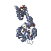
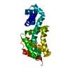
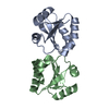
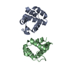
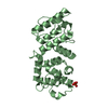
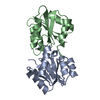
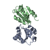
| Title | Crystal structure of dimeric K72A human cytochrome c alkaline conformer | ||||||
 Components Components | Cytochrome c | ||||||
 Keywords Keywords | ELECTRON TRANSPORT / cytochrome c / cardiolipin / heme protein | ||||||
| Function / homology |  Function and homology information Function and homology informationFormation of apoptosome / apoptosome / activation of cysteine-type endopeptidase activity involved in apoptotic process by cytochrome c / Release of apoptotic factors from the mitochondria / Respiratory electron transport / cellular respiration / Regulation of the apoptosome activity / Activation of caspases through apoptosome-mediated cleavage / SMAC (DIABLO) binds to IAPs / SMAC(DIABLO)-mediated dissociation of IAP:caspase complexes ...Formation of apoptosome / apoptosome / activation of cysteine-type endopeptidase activity involved in apoptotic process by cytochrome c / Release of apoptotic factors from the mitochondria / Respiratory electron transport / cellular respiration / Regulation of the apoptosome activity / Activation of caspases through apoptosome-mediated cleavage / SMAC (DIABLO) binds to IAPs / SMAC(DIABLO)-mediated dissociation of IAP:caspase complexes / mitochondrial electron transport, cytochrome c to oxygen / mitochondrial electron transport, ubiquinol to cytochrome c / Detoxification of Reactive Oxygen Species / Pyroptosis / respirasome / intrinsic apoptotic signaling pathway / TP53 Regulates Metabolic Genes / Transcriptional activation of mitochondrial biogenesis / mitochondrial intermembrane space / Cytoprotection by HMOX1 / mitochondrial inner membrane / electron transfer activity / heme binding / mitochondrion / metal ion binding / nucleus / cytosolSimilarity search - Function | ||||||
| Biological species |  Homo sapiens (human) Homo sapiens (human) | ||||||
| Method | X-RAY DIFFRACTION / SYNCHROTRON / MOLECULAR REPLACEMENT / Resolution: 2.08 Å | ||||||
 Authors Authors | Lei, H. / Bowler, B.E. | ||||||
| Funding support |  United States, 1items United States, 1items
| ||||||
 Citation Citation | Journal: J.Am.Chem.Soc. / Year: 2022 Title: Alkaline State of the Domain-Swapped Dimer of Human Cytochrome c : A Conformational Switch for Apoptotic Peroxidase Activity. Authors: Lei, H. / Kelly, A.D. / Bowler, B.E. | ||||||
| History |
|
- Structure visualization
| Structure viewer | Molecule: MolmilJmol/JSmol |
|---|
- Downloads & links
-Download
| PDBx/mmCIF format | 6xnk.cif.gz | 213.6 KB | Display | PDBx/mmCIF format |
|---|---|---|---|---|
| PDB format | pdb6xnk.ent.gz | 140.7 KB | Display | PDB format |
| PDBx/mmJSON format | 6xnk.json.gz | Tree view | PDBx/mmJSON format | |
| Others |  Other downloads Other downloads |
-Validation report
| Arichive directory | https://data.pdbj.org/pub/pdb/validation_reports/xn/6xnkftp://data.pdbj.org/pub/pdb/validation_reports/xn/6xnk | HTTPS FTP |
|---|
-Related structure data
| Related structure data |  5ty3S S: Starting model for refinement |
|---|---|
| Similar structure data |


-Links
 PDBj
PDBj


- Assembly
| Deposited unit | 
| ||||||||||||||||||||||||||||||
|---|---|---|---|---|---|---|---|---|---|---|---|---|---|---|---|---|---|---|---|---|---|---|---|---|---|---|---|---|---|---|---|
| 1 | 
| ||||||||||||||||||||||||||||||
| 2 | 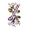
| ||||||||||||||||||||||||||||||
| Unit cell |
| ||||||||||||||||||||||||||||||
| Noncrystallographic symmetry (NCS) | NCS domain:
NCS domain segments: Component-ID: 1 / Ens-ID: 1 / Beg auth comp-ID: GLY / Beg label comp-ID: GLY / End auth comp-ID: GLU / End label comp-ID: GLU / Auth seq-ID: 1 - 104 / Label seq-ID: 1 - 104
|
-Components
| #1: Protein | Mass: 11582.479 Da / Num. of mol.: 4 / Mutation: K72A Source method: isolated from a genetically manipulated source Source: (gene. exp.) Homo sapiens (human) / Gene: CYCS, CYC / Production host:  Escherichia coli (E. coli) / References: UniProt: P99999 Escherichia coli (E. coli) / References: UniProt: P99999#2: Chemical | ChemComp-HEC / Heme C  Mass: 618.503 Da / Num. of mol.: 4 / Source method: obtained synthetically / Formula: C34H34FeN4O4 / Feature type: SUBJECT OF INVESTIGATION Mass: 618.503 Da / Num. of mol.: 4 / Source method: obtained synthetically / Formula: C34H34FeN4O4 / Feature type: SUBJECT OF INVESTIGATION#3: Water | ChemComp-HOH / | Water Mass: 18.015 Da / Num. of mol.: 109 / Source method: isolated from a natural source / Formula: H2O Mass: 18.015 Da / Num. of mol.: 109 / Source method: isolated from a natural source / Formula: H2OHas ligand of interest | Y | |
|---|
-Experimental details
-Experiment
| Experiment | Method: X-RAY DIFFRACTION / Number of used crystals: 1 |
|---|
- Sample preparation
| Crystal | Density Matthews: 2.25 Å3/Da / Density % sol: 45.43 % |
|---|---|
| Crystal grow | Temperature: 293 K / Method: vapor diffusion, sitting drop / pH: 9.9 / Details: 30% MPD, 0.1 M CHES pH 9.9 / PH range: 9-9.9 |
-Data collection
| Diffraction | Mean temperature: 100 K / Serial crystal experiment: N |
|---|---|
| Diffraction source | Source: SYNCHROTRON / Site: APS / Beamline: 19-ID / Wavelength: 0.987 Å |
| Detector | Type: DECTRIS PILATUS3 X 6M / Detector: PIXEL / Date: Feb 18, 2018 |
| Radiation | Protocol: SINGLE WAVELENGTH / Monochromatic (M) / Laue (L): M / Scattering type: x-ray |
| Radiation wavelength | Wavelength: 0.987 Å / Relative weight: 1 |
| Reflection | Resolution: 2.08→42.12 Å / Num. obs: 22075 / % possible obs: 90.5 % / Redundancy: 3 % / Biso Wilson estimate: 28.4888322735 Å2 / Rmerge(I) obs: 0.14 / Net I/σ(I): 3.05 |
| Reflection shell | Resolution: 2.08→2.15 Å / Rmerge(I) obs: 0.6 / Num. unique obs: 1757 / % possible all: 72.4 |
- Processing
| Software |
| |||||||||||||||||||||||||||||||||||||||||||||||||||||||||||||||||||||||||||||||||||||||||||||||||||||||||
|---|---|---|---|---|---|---|---|---|---|---|---|---|---|---|---|---|---|---|---|---|---|---|---|---|---|---|---|---|---|---|---|---|---|---|---|---|---|---|---|---|---|---|---|---|---|---|---|---|---|---|---|---|---|---|---|---|---|---|---|---|---|---|---|---|---|---|---|---|---|---|---|---|---|---|---|---|---|---|---|---|---|---|---|---|---|---|---|---|---|---|---|---|---|---|---|---|---|---|---|---|---|---|---|---|---|---|
| Refinement | Method to determine structure: MOLECULAR REPLACEMENT Starting model: 5ty3 Resolution: 2.08→42.11 Å / SU ML: 0.25 / Cross valid method: FREE R-VALUE / σ(F): 1.96 / Phase error: 25.48
| |||||||||||||||||||||||||||||||||||||||||||||||||||||||||||||||||||||||||||||||||||||||||||||||||||||||||
| Solvent computation | Shrinkage radii: 0.9 Å / VDW probe radii: 1.11 Å | |||||||||||||||||||||||||||||||||||||||||||||||||||||||||||||||||||||||||||||||||||||||||||||||||||||||||
| Displacement parameters | Biso mean: 42.9 Å2 | |||||||||||||||||||||||||||||||||||||||||||||||||||||||||||||||||||||||||||||||||||||||||||||||||||||||||
| Refinement step | Cycle: LAST / Resolution: 2.08→42.11 Å
| |||||||||||||||||||||||||||||||||||||||||||||||||||||||||||||||||||||||||||||||||||||||||||||||||||||||||
| Refine LS restraints |
| |||||||||||||||||||||||||||||||||||||||||||||||||||||||||||||||||||||||||||||||||||||||||||||||||||||||||
| LS refinement shell |
|
