 Movie
Movie Controller

[English] 日本語
 Yorodumi
- PDB-5g2z: Crystallographic structure of mutant W31A of thioredoxin from Lit... -
+ Open data

- Basic information
| Entry | Database: PDB / ID: 5g2z | ||||||
|---|---|---|---|---|---|---|---|
| Title | Crystallographic structure of mutant W31A of thioredoxin from Litopenaeus vannamei | ||||||
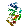
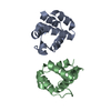
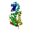
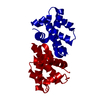
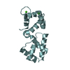
 Components Components | THIOREDOXIN | ||||||
 Keywords Keywords | OXIDOREDUCTASE / THIOREDOXIN / SHRIMP / LITOPENAEUS VANNAMEI / MUTANT / DISULFIDE BOND | ||||||
| Function / homology |  Function and homology information Function and homology information | ||||||
| Biological species |  LITOPENAEUS VANNAMEI (Pacific white shrimp) LITOPENAEUS VANNAMEI (Pacific white shrimp) | ||||||
| Method | X-RAY DIFFRACTION / MOLECULAR REPLACEMENT / Resolution: 1.88 Å | ||||||
 Authors Authors | Campos-Acevedo, A.A. / Rudino-Pinera, E. | ||||||
 Citation Citation | Journal: Acta Crystallogr D Struct Biol / Year: 2017 Title: Is dimerization a common feature in thioredoxins? The case of thioredoxin from Litopenaeus vannamei. Authors: Campos-Acevedo, A.A. / Sotelo-Mundo, R.R. / Perez, J. / Rudino-Pinera, E. | ||||||
| History |
|
- Structure visualization
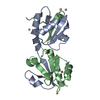
| Structure viewer | Molecule: MolmilJmol/JSmol |
|---|
- Downloads & links
-Download
| PDBx/mmCIF format | 5g2z.cif.gz | 55.2 KB | Display | PDBx/mmCIF format |
|---|---|---|---|---|
| PDB format | pdb5g2z.ent.gz | 40.8 KB | Display | PDB format |
| PDBx/mmJSON format | 5g2z.json.gz | Tree view | PDBx/mmJSON format | |
| Others |  Other downloads Other downloads |
-Validation report
| Arichive directory | https://data.pdbj.org/pub/pdb/validation_reports/g2/5g2zftp://data.pdbj.org/pub/pdb/validation_reports/g2/5g2z | HTTPS FTP |
|---|
-Related structure data
| Related structure data |  5g30C  5g31C 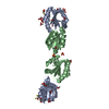 3zzxS C: citing same article ( S: Starting model for refinement |
|---|---|
| Similar structure data |


-Links
 PDBj
PDBj

- Assembly
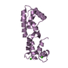
| Deposited unit | 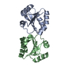
| ||||||||
|---|---|---|---|---|---|---|---|---|---|
| 1 |
| ||||||||
| Unit cell |
|
-Components
| #1: Protein | Mass: 11843.578 Da / Num. of mol.: 2 / Mutation: YES Source method: isolated from a genetically manipulated source Details: RESIDUE 11 IS A SER IN THE UNIPROT DEPOSIT B1PWB9, IN THIS STRUCTURE A PHE IS CLEARLY VISIBLE ON POSITION 11. Source: (gene. exp.) LITOPENAEUS VANNAMEI (Pacific white shrimp)Plasmid: PET22B / Production host:  ESCHERICHIA COLI (E. coli) ESCHERICHIA COLI (E. coli)References: UniProt: B1PWB9, thioredoxin-disulfide reductase#2: Water | ChemComp-HOH / | Water Mass: 18.015 Da / Num. of mol.: 140 / Source method: isolated from a natural source / Formula: H2O Mass: 18.015 Da / Num. of mol.: 140 / Source method: isolated from a natural source / Formula: H2OSequence details | A SERINE IS PRESENT IN RESIDUE 11 FROM UNIPROT DEPOSIT B1PWB9, IN OUR STRUCTURE RESIDUE 11 IS A PHENILALAN | |
|---|
-Experimental details
-Experiment
| Experiment | Method: X-RAY DIFFRACTION / Number of used crystals: 1 |
|---|
- Sample preparation
| Crystal | Density Matthews: 2.15 Å3/Da / Density % sol: 42.78 % / Description: NONE |
|---|---|
| Crystal grow | pH: 7 Details: 30% (W/V) POLYETHYLENE GLYCOL 3, 000. 100 MM TRIS BASE/HYDROCHLORIC ACID PH 7.0. 200 MM NACL |
-Data collection
| Diffraction | Mean temperature: 100 K |
|---|---|
| Diffraction source | Source: ROTATING ANODE / Wavelength: 1.5418 |
| Detector | Type: RIGAKU-MSC R-AXIS IV / Detector: IMAGE PLATE / Date: Nov 18, 2014 / Details: VARIMAX-HR |
| Radiation | Protocol: SINGLE WAVELENGTH / Monochromatic (M) / Laue (L): M / Scattering type: x-ray |
| Radiation wavelength | Wavelength: 1.5418 Å / Relative weight: 1 |
| Reflection | Resolution: 1.88→23.43 Å / Num. obs: 17415 / % possible obs: 99.4 % / Observed criterion σ(I): 0 / Redundancy: 4 % / Biso Wilson estimate: 23.21 Å2 / Rmerge(I) obs: 0.04 / Net I/σ(I): 17.6 |
| Reflection shell | Resolution: 1.88→1.92 Å / Redundancy: 3.9 % / Rmerge(I) obs: 0.21 / Mean I/σ(I) obs: 4.6 / % possible all: 100 |
- Processing
| Software |
| |||||||||||||||||||||||||||||||||||||||||||||||||||||||||||||||||||||||||||||||||||||||||||
|---|---|---|---|---|---|---|---|---|---|---|---|---|---|---|---|---|---|---|---|---|---|---|---|---|---|---|---|---|---|---|---|---|---|---|---|---|---|---|---|---|---|---|---|---|---|---|---|---|---|---|---|---|---|---|---|---|---|---|---|---|---|---|---|---|---|---|---|---|---|---|---|---|---|---|---|---|---|---|---|---|---|---|---|---|---|---|---|---|---|---|---|---|
| Refinement | Method to determine structure: MOLECULAR REPLACEMENT Starting model: PDB ENTRY 3ZZX Resolution: 1.88→23.429 Å / SU ML: 0.2 / σ(F): 1.36 / Phase error: 22.19 / Stereochemistry target values: ML
| |||||||||||||||||||||||||||||||||||||||||||||||||||||||||||||||||||||||||||||||||||||||||||
| Solvent computation | Shrinkage radii: 0.9 Å / VDW probe radii: 1.11 Å / Solvent model: FLAT BULK SOLVENT MODEL | |||||||||||||||||||||||||||||||||||||||||||||||||||||||||||||||||||||||||||||||||||||||||||
| Displacement parameters | Biso mean: 28.26 Å2 | |||||||||||||||||||||||||||||||||||||||||||||||||||||||||||||||||||||||||||||||||||||||||||
| Refinement step | Cycle: LAST / Resolution: 1.88→23.429 Å
| |||||||||||||||||||||||||||||||||||||||||||||||||||||||||||||||||||||||||||||||||||||||||||
| Refine LS restraints |
| |||||||||||||||||||||||||||||||||||||||||||||||||||||||||||||||||||||||||||||||||||||||||||
| LS refinement shell |
|
