 Movie
Movie Controller


+ Open data

- Basic information
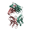
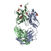
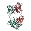
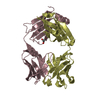
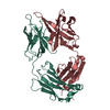
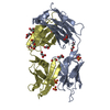
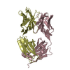
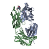
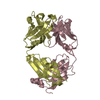
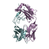
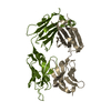
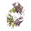
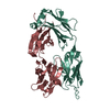
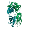
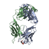
| Entry | Database: PDB / ID: 6x5e | ||||||
|---|---|---|---|---|---|---|---|
| Title | Crystal structure of a Lewis-binding Fab (ch88.2) | ||||||
 Components Components |
| ||||||
 Keywords Keywords |  IMMUNE SYSTEM / immunoglobulin / chimeric antibody / antigen binding fragment / Lewis-binding antibody IMMUNE SYSTEM / immunoglobulin / chimeric antibody / antigen binding fragment / Lewis-binding antibody | ||||||
| Function / homology | NICKEL (II) ION Function and homology information Function and homology information | ||||||
| Biological species |  Mus musculus (house mouse) Mus musculus (house mouse) | ||||||
| Method | X-RAY DIFFRACTION / SYNCHROTRON / MOLECULAR REPLACEMENT / Resolution: 2.29 Å | ||||||
 Authors Authors | Soliman, C. / Ramsland, P.A. | ||||||
 Citation Citation | Journal: Biochem.J. / Year: 2020 Title: Molecular and structural basis for Lewis glycan recognition by a cancer-targeting antibody. Authors: Soliman, C. / Guy, A.J. / Chua, J.X. / Vankemmelbeke, M. / McIntosh, R.S. / Eastwood, S. / Truong, V.K. / Elbourne, A. / Spendlove, I. / Durrant, L.G. / Ramsland, P.A. | ||||||
| History |
|
- Structure visualization
| Structure viewer | Molecule: MolmilJmol/JSmol |
|---|
- Downloads & links
-Download
| PDBx/mmCIF format | 6x5e.cif.gz | 392.3 KB | Display | PDBx/mmCIF format |
|---|---|---|---|---|
| PDB format | pdb6x5e.ent.gz | 261.4 KB | Display | PDB format |
| PDBx/mmJSON format | 6x5e.json.gz | Tree view | PDBx/mmJSON format | |
| Others |  Other downloads Other downloads |
-Validation report
| Arichive directory | https://data.pdbj.org/pub/pdb/validation_reports/x5/6x5eftp://data.pdbj.org/pub/pdb/validation_reports/x5/6x5e | HTTPS FTP |
|---|
-Related structure data
| Related structure data |  4x80S S: Starting model for refinement |
|---|---|
| Similar structure data |


-Links
 PDBj
PDBj

- Assembly
| Deposited unit | 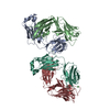
| ||||||||||
|---|---|---|---|---|---|---|---|---|---|---|---|
| 1 | 
| ||||||||||
| 2 | 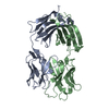
| ||||||||||
| Unit cell |
|
-Components
| #1: Antibody | Mass: 23644.180 Da / Num. of mol.: 2 Source method: isolated from a genetically manipulated source Source: (gene. exp.) Mus musculus (house mouse) / Production host:  Homo sapiens (human) Homo sapiens (human)#2: Antibody | Mass: 24431.475 Da / Num. of mol.: 2 Source method: isolated from a genetically manipulated source Source: (gene. exp.) Mus musculus (house mouse) / Production host: Homo sapiens (human)#3: Chemical | Nickel  Mass: 58.693 Da / Num. of mol.: 3 / Source method: obtained synthetically / Formula: Ni Mass: 58.693 Da / Num. of mol.: 3 / Source method: obtained synthetically / Formula: Ni#4: Water | ChemComp-HOH / | Water Mass: 18.015 Da / Num. of mol.: 36 / Source method: isolated from a natural source / Formula: H2O Mass: 18.015 Da / Num. of mol.: 36 / Source method: isolated from a natural source / Formula: H2OHas ligand of interest | N | |
|---|
-Experimental details
-Experiment
| Experiment | Method: X-RAY DIFFRACTION / Number of used crystals: 1 |
|---|
- Sample preparation
| Crystal | Density Matthews: 2.3 Å3/Da / Density % sol: 47.2 % |
|---|---|
| Crystal grow | Temperature: 291 K / Method: vapor diffusion, sitting drop / pH: 8.5 / Details: PEG MME 200, Tris, Nickel (II) chloride |
-Data collection
| Diffraction | Mean temperature: 100 K / Serial crystal experiment: N |
|---|---|
| Diffraction source | Source: SYNCHROTRON / Site: Australian Synchrotron  / Beamline: MX2 / Wavelength: 0.954 Å / Beamline: MX2 / Wavelength: 0.954 Å |
| Detector | Type: DECTRIS EIGER X 16M / Detector: PIXEL / Date: Jun 29, 2019 |
| Radiation | Protocol: SINGLE WAVELENGTH / Monochromatic (M) / Laue (L): M / Scattering type: x-ray |
| Radiation wavelength | Wavelength: 0.954 Å / Relative weight: 1 |
| Reflection | Resolution: 2.29→50 Å / Num. obs: 44428 / % possible obs: 92.6 % / Redundancy: 1.8 % / Biso Wilson estimate: 60.41 Å2 / CC1/2: 0.998 / Rrim(I) all: 0.07 / Net I/σ(I): 7.9 |
| Reflection shell | Resolution: 2.29→2.4 Å / Num. unique obs: 6787 / CC1/2: 0.719 / Rrim(I) all: 0.88 |
- Processing
| Software |
| |||||||||||||||||||||||||||||||||||||||||||||||||||||||||||||||||||||||||||||||||||||||||||||||||||||||||
|---|---|---|---|---|---|---|---|---|---|---|---|---|---|---|---|---|---|---|---|---|---|---|---|---|---|---|---|---|---|---|---|---|---|---|---|---|---|---|---|---|---|---|---|---|---|---|---|---|---|---|---|---|---|---|---|---|---|---|---|---|---|---|---|---|---|---|---|---|---|---|---|---|---|---|---|---|---|---|---|---|---|---|---|---|---|---|---|---|---|---|---|---|---|---|---|---|---|---|---|---|---|---|---|---|---|---|
| Refinement | Method to determine structure: MOLECULAR REPLACEMENT Starting model: 4X80 Resolution: 2.29→33.49 Å / SU ML: 0.4361 / Cross valid method: FREE R-VALUE / σ(F): 1.87 / Phase error: 37.6612 Stereochemistry target values: GeoStd + Monomer Library + CDL v1.2
| |||||||||||||||||||||||||||||||||||||||||||||||||||||||||||||||||||||||||||||||||||||||||||||||||||||||||
| Solvent computation | Shrinkage radii: 0.9 Å / VDW probe radii: 1.11 Å / Solvent model: FLAT BULK SOLVENT MODEL | |||||||||||||||||||||||||||||||||||||||||||||||||||||||||||||||||||||||||||||||||||||||||||||||||||||||||
| Displacement parameters | Biso mean: 68.4 Å2 | |||||||||||||||||||||||||||||||||||||||||||||||||||||||||||||||||||||||||||||||||||||||||||||||||||||||||
| Refinement step | Cycle: LAST / Resolution: 2.29→33.49 Å
| |||||||||||||||||||||||||||||||||||||||||||||||||||||||||||||||||||||||||||||||||||||||||||||||||||||||||
| Refine LS restraints |
| |||||||||||||||||||||||||||||||||||||||||||||||||||||||||||||||||||||||||||||||||||||||||||||||||||||||||
| LS refinement shell |
|
