 Movie
Movie Controller


+ Open data

- Basic information
| Entry | Database: PDB / ID: 6w65 | ||||||
|---|---|---|---|---|---|---|---|
| Title | Human PARP16 in complex with RBN010860 | ||||||
 Components Components | Protein mono-ADP-ribosyltransferase PARP16 | ||||||
 Keywords Keywords |  TRANSFERASE / ADP-RIBOSE / PARP16 / ARTD15 / ARTD Transferase domain / ADP-ribosylation / TRANSFERASE-TRANSFERASE INHIBITOR complex TRANSFERASE / ADP-RIBOSE / PARP16 / ARTD15 / ARTD Transferase domain / ADP-ribosylation / TRANSFERASE-TRANSFERASE INHIBITOR complex | ||||||
| Function / homology |  Function and homology information Function and homology informationendoplasmic reticulum tubular network / NAD+- protein-lysine ADP-ribosyltransferase activity / NAD biosynthesis via nicotinamide riboside salvage pathway / NAD+- protein-aspartate ADP-ribosyltransferase activity / NAD+-protein-glutamate ADP-ribosyltransferase activity / Nicotinamide salvaging / Maturation of nucleoprotein / Maturation of nucleoprotein / protein auto-ADP-ribosylation / IRE1-mediated unfolded protein response ...endoplasmic reticulum tubular network / NAD+- protein-lysine ADP-ribosyltransferase activity / NAD biosynthesis via nicotinamide riboside salvage pathway / NAD+- protein-aspartate ADP-ribosyltransferase activity / NAD+-protein-glutamate ADP-ribosyltransferase activity / Nicotinamide salvaging / Maturation of nucleoprotein / Maturation of nucleoprotein / protein auto-ADP-ribosylation / IRE1-mediated unfolded protein response / NAD+-protein ADP-ribosyltransferase activity / Transferases; Glycosyltransferases; Pentosyltransferases / NAD+ ADP-ribosyltransferase activity / negative regulation of cytoplasmic translation / endoplasmic reticulum unfolded protein response / nucleotidyltransferase activity / protein serine/threonine kinase activator activity / cellular response to leukemia inhibitory factor / kinase binding / nuclear envelope / viral protein processing / endoplasmic reticulum membrane / endoplasmic reticulum / membrane / cytosolSimilarity search - Function | ||||||
| Biological species |  Homo sapiens (human) Homo sapiens (human) | ||||||
| Method | X-RAY DIFFRACTION / SYNCHROTRON / MOLECULAR REPLACEMENT / Resolution: 2.13 Å | ||||||
 Authors Authors | Swinger, K.K. / Wigle, T.J. / Kuntz, K.W. | ||||||
 Citation Citation | Journal: Cell Chem Biol / Year: 2020 Title: In Vitro and Cellular Probes to Study PARP Enzyme Target Engagement. Authors: Wigle, T.J. / Blackwell, D.J. / Schenkel, L.B. / Ren, Y. / Church, W.D. / Desai, H.J. / Swinger, K.K. / Santospago, A.G. / Majer, C.R. / Lu, A.Z. / Niepel, M. / Perl, N.R. / Vasbinder, M.M. ...Authors: Wigle, T.J. / Blackwell, D.J. / Schenkel, L.B. / Ren, Y. / Church, W.D. / Desai, H.J. / Swinger, K.K. / Santospago, A.G. / Majer, C.R. / Lu, A.Z. / Niepel, M. / Perl, N.R. / Vasbinder, M.M. / Keilhack, H. / Kuntz, K.W. | ||||||
| History |
|
- Structure visualization
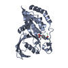
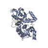
| Structure viewer | Molecule: MolmilJmol/JSmol |
|---|
- Downloads & links
-Download
| PDBx/mmCIF format | 6w65.cif.gz | 168.7 KB | Display | PDBx/mmCIF format |
|---|---|---|---|---|
| PDB format | pdb6w65.ent.gz | 133.1 KB | Display | PDB format |
| PDBx/mmJSON format | 6w65.json.gz | Tree view | PDBx/mmJSON format | |
| Others |  Other downloads Other downloads |
-Validation report
| Arichive directory | https://data.pdbj.org/pub/pdb/validation_reports/w6/6w65ftp://data.pdbj.org/pub/pdb/validation_reports/w6/6w65 | HTTPS FTP |
|---|
-Related structure data
| Related structure data | |
|---|---|
| Similar structure data |


-Links
 PDBj
PDBj- Assembly
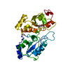
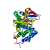
| Deposited unit | 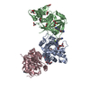
| |||||||||||||||||||||||||||||||||||||||||||||||||||||
|---|---|---|---|---|---|---|---|---|---|---|---|---|---|---|---|---|---|---|---|---|---|---|---|---|---|---|---|---|---|---|---|---|---|---|---|---|---|---|---|---|---|---|---|---|---|---|---|---|---|---|---|---|---|---|
| 1 | 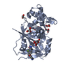
| |||||||||||||||||||||||||||||||||||||||||||||||||||||
| 2 | 
| |||||||||||||||||||||||||||||||||||||||||||||||||||||
| 3 | 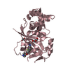
| |||||||||||||||||||||||||||||||||||||||||||||||||||||
| Unit cell |
| |||||||||||||||||||||||||||||||||||||||||||||||||||||
| Components on special symmetry positions |
| |||||||||||||||||||||||||||||||||||||||||||||||||||||
| Noncrystallographic symmetry (NCS) | NCS domain:
NCS domain segments: Component-ID: 0 / Beg auth comp-ID: SER / Beg label comp-ID: SER / End auth comp-ID: LYS / End label comp-ID: LYS / Refine code: 0 / Auth seq-ID: 3 - 274 / Label seq-ID: 1 - 272
NCS ensembles :
|
-Components
-Protein , 1 types, 3 molecules ABC
| #1: Protein | Mass: 31110.578 Da / Num. of mol.: 3 Source method: isolated from a genetically manipulated source Source: (gene. exp.) Homo sapiens (human) / Gene: PARP16, ARTD15, C15orf30 / Production host:  Escherichia coli (E. coli) Escherichia coli (E. coli)References: UniProt: Q8N5Y8, Transferases; Glycosyltransferases; Pentosyltransferases |
|---|
-Non-polymers , 6 types, 300 molecules 


| #2: Chemical |  Mass: 378.348 Da / Num. of mol.: 3 / Source method: obtained synthetically / Formula: C18H17F3N4O2 / Feature type: SUBJECT OF INVESTIGATION Mass: 378.348 Da / Num. of mol.: 3 / Source method: obtained synthetically / Formula: C18H17F3N4O2 / Feature type: SUBJECT OF INVESTIGATION#3: Chemical | Citric acid Mass: 192.124 Da / Num. of mol.: 2 / Source method: obtained synthetically / Formula: C6H8O7 Mass: 192.124 Da / Num. of mol.: 2 / Source method: obtained synthetically / Formula: C6H8O7#4: Chemical | ChemComp-GOL / Glycerol Mass: 92.094 Da / Num. of mol.: 10 / Source method: obtained synthetically / Formula: C3H8O3 Mass: 92.094 Da / Num. of mol.: 10 / Source method: obtained synthetically / Formula: C3H8O3#5: Chemical | ChemComp-EDO / Ethylene glycol Mass: 62.068 Da / Num. of mol.: 4 / Source method: obtained synthetically / Formula: C2H6O2 Mass: 62.068 Da / Num. of mol.: 4 / Source method: obtained synthetically / Formula: C2H6O2#6: Chemical | Diethylene glycol Mass: 106.120 Da / Num. of mol.: 2 / Source method: obtained synthetically / Formula: C4H10O3 Mass: 106.120 Da / Num. of mol.: 2 / Source method: obtained synthetically / Formula: C4H10O3#7: Water | ChemComp-HOH / | WaterMass: 18.015 Da / Num. of mol.: 279 / Source method: isolated from a natural source / Formula: H2O |
|---|
-Details
| Has ligand of interest | Y |
|---|
-Experimental details
-Experiment
| Experiment | Method: X-RAY DIFFRACTION / Number of used crystals: 1 |
|---|
- Sample preparation
| Crystal | Density Matthews: 2.88 Å3/Da / Density % sol: 57.33 % |
|---|---|
| Crystal grow | Temperature: 291.15 K / Method: vapor diffusion, sitting drop / Details: 100mM Sodium citrate pH5.5, 18%w/v PEG3350 |
-Data collection
| Diffraction | Mean temperature: 63 K / Serial crystal experiment: N |
|---|---|
| Diffraction source | Source: SYNCHROTRON / Site: SSRF  / Beamline: BL17U1 / Wavelength: 0.9792 Å / Beamline: BL17U1 / Wavelength: 0.9792 Å |
| Detector | Type: DECTRIS EIGER2 X 9M / Detector: PIXEL / Date: Oct 28, 2019 |
| Radiation | Protocol: SINGLE WAVELENGTH / Monochromatic (M) / Laue (L): M / Scattering type: x-ray |
| Radiation wavelength | Wavelength: 0.9792 Å / Relative weight: 1 |
| Reflection | Resolution: 2.13→37.78 Å / Num. obs: 58280 / % possible obs: 99.87 % / Redundancy: 12.5 % / Rmerge(I) obs: 0.061 / Net I/σ(I): 23.6 |
| Reflection shell | Resolution: 2.13→2.185 Å / Rmerge(I) obs: 0.948 / Num. unique obs: 4242 |
- Processing
| Software |
| ||||||||||||||||||||||||||||||||||||||||||||||||||||||||||||
|---|---|---|---|---|---|---|---|---|---|---|---|---|---|---|---|---|---|---|---|---|---|---|---|---|---|---|---|---|---|---|---|---|---|---|---|---|---|---|---|---|---|---|---|---|---|---|---|---|---|---|---|---|---|---|---|---|---|---|---|---|---|
| Refinement | Method to determine structure: MOLECULAR REPLACEMENT / Resolution: 2.13→37.78 Å / Cor.coef. Fo:Fc: 0.962 / Cor.coef. Fo:Fc free: 0.944 / SU B: 4.942 / SU ML: 0.124 / Cross valid method: THROUGHOUT / σ(F): 0 / ESU R: 0.177 / ESU R Free: 0.159 Details: HYDROGENS HAVE BEEN ADDED IN THE RIDING POSITIONS U VALUES : REFINED INDIVIDUALLY
| ||||||||||||||||||||||||||||||||||||||||||||||||||||||||||||
| Solvent computation | Ion probe radii: 0.8 Å / Shrinkage radii: 0.8 Å / VDW probe radii: 1.2 Å | ||||||||||||||||||||||||||||||||||||||||||||||||||||||||||||
| Displacement parameters | Biso max: 158.68 Å2 / Biso mean: 55.531 Å2 / Biso min: 26.67 Å2
| ||||||||||||||||||||||||||||||||||||||||||||||||||||||||||||
| Refinement step | Cycle: final / Resolution: 2.13→37.78 Å
| ||||||||||||||||||||||||||||||||||||||||||||||||||||||||||||
| Refine LS restraints |
| ||||||||||||||||||||||||||||||||||||||||||||||||||||||||||||
| Refine LS restraints NCS | Refine-ID: X-RAY DIFFRACTION / Type: interatomic distance / Weight position: 0.05
| ||||||||||||||||||||||||||||||||||||||||||||||||||||||||||||
| LS refinement shell | Resolution: 2.13→2.185 Å / Rfactor Rfree error: 0 / Total num. of bins used: 20
|
