 Movie
Movie Controller


+ Open data

- Basic information
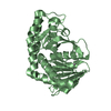
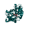
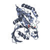
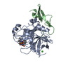
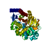
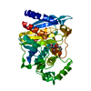
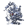
| Entry | Database: PDB / ID: 5via | ||||||
|---|---|---|---|---|---|---|---|
| Title | Crystal structural of Leishmania major pseudoperoxidase | ||||||
 Components Components | Pseudoperoxidase | ||||||
 Keywords Keywords |  OXIDOREDUCTASE / heme protein / peroxidase OXIDOREDUCTASE / heme protein / peroxidase | ||||||
| Function / homology |  Function and homology information Function and homology informationresponse to reactive oxygen species / hydrogen peroxide catabolic process / peroxidase activity / cellular response to oxidative stress / membrane => GO:0016020 / heme binding / metal ion bindingSimilarity search - Function | ||||||
| Biological species |  Leishmania major (eukaryote) Leishmania major (eukaryote) | ||||||
| Method | X-RAY DIFFRACTION / SYNCHROTRON / MAD / Resolution: 1.764 Å | ||||||
 Authors Authors | Chreifi, G. / Dejam, D. / Poulos, T.L. | ||||||
| Funding support |  United States, 1items United States, 1items
| ||||||
 Citation Citation | Journal: J. Biol. Inorg. Chem. / Year: 2017 Title: Crystal structure and functional analysis of Leishmania major pseudoperoxidase. Authors: Chreifi, G. / Dejam, D. / Poulos, T.L. | ||||||
| History |
|
- Structure visualization
| Structure viewer | Molecule: MolmilJmol/JSmol |
|---|
- Downloads & links
-Download
| PDBx/mmCIF format | 5via.cif.gz | 129.1 KB | Display | PDBx/mmCIF format |
|---|---|---|---|---|
| PDB format | pdb5via.ent.gz | 100 KB | Display | PDB format |
| PDBx/mmJSON format | 5via.json.gz | Tree view | PDBx/mmJSON format | |
| Others |  Other downloads Other downloads |
-Validation report
| Arichive directory | https://data.pdbj.org/pub/pdb/validation_reports/vi/5viaftp://data.pdbj.org/pub/pdb/validation_reports/vi/5via | HTTPS FTP |
|---|
-Related structure data
| Similar structure data |
|---|


-Links
 PDBj
PDBj
- Assembly
| Deposited unit | 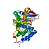
| ||||||||
|---|---|---|---|---|---|---|---|---|---|
| 1 |
| ||||||||
| Unit cell |
| ||||||||
| Components on special symmetry positions |
|
-Components
| #1: Protein | Mass: 33066.293 Da / Num. of mol.: 1 / Fragment: UNP residues 49-341 Source method: isolated from a genetically manipulated source Source: (gene. exp.) Leishmania major (eukaryote) / Gene: LMJF_21_1567 / Production host:  Escherichia coli BL21(DE3) (bacteria) / References: UniProt: Q4QC30 Escherichia coli BL21(DE3) (bacteria) / References: UniProt: Q4QC30 |
|---|---|
| #2: Chemical | ChemComp-HEM / Heme B  Mass: 616.487 Da / Num. of mol.: 1 / Source method: obtained synthetically / Formula: C34H32FeN4O4 Mass: 616.487 Da / Num. of mol.: 1 / Source method: obtained synthetically / Formula: C34H32FeN4O4 |
| #3: Water | ChemComp-HOH / Water Mass: 18.015 Da / Num. of mol.: 181 / Source method: isolated from a natural source / Formula: H2O Mass: 18.015 Da / Num. of mol.: 181 / Source method: isolated from a natural source / Formula: H2O |
-Experimental details
-Experiment
| Experiment | Method: X-RAY DIFFRACTION / Number of used crystals: 1 |
|---|
- Sample preparation
| Crystal | Density Matthews: 2.34 Å3/Da / Density % sol: 47.38 % |
|---|---|
| Crystal grow | Temperature: 296 K / Method: vapor diffusion, hanging drop / pH: 7.5 Details: 14 mg/mL protein, 5% 2-methyl-2,4-pentanediol, 10% polyethylene glycol 6000, and 0.1 M HEPES pH 7.5 |
-Data collection
| Diffraction | Mean temperature: 100 K |
|---|---|
| Diffraction source | Source: SYNCHROTRON / Site: SSRL / Beamline: BL14-1 / Wavelength: 1.18076 Å |
| Detector | Type: RAYONIX MX325HE / Detector: CCD / Date: Dec 16, 2015 |
| Radiation | Protocol: MAD / Monochromatic (M) / Laue (L): M / Scattering type: x-ray |
| Radiation wavelength | Wavelength: 1.18076 Å / Relative weight: 1 |
| Reflection | Resolution: 1.76→48.88 Å / Num. obs: 31236 / % possible obs: 98.22 % / Redundancy: 18 % / Biso Wilson estimate: 34.91 Å2 / CC1/2: 0.998 / Rmerge(I) obs: 0.086 / Rpim(I) all: 0.049 / Net I/σ(I): 73.11 |
| Reflection shell | Resolution: 1.76→1.83 Å / Redundancy: 16.5 % / Num. unique obs: 3100 / CC1/2: 0.297 / % possible all: 94.03 |
- Processing
| Software |
| |||||||||||||||||||||||||||||||||||||||||||||||||||||||||||||||||||||||||||||||||||||||||||||||||||||||||||||||||||||||||||||||||||||||||||||||||||
|---|---|---|---|---|---|---|---|---|---|---|---|---|---|---|---|---|---|---|---|---|---|---|---|---|---|---|---|---|---|---|---|---|---|---|---|---|---|---|---|---|---|---|---|---|---|---|---|---|---|---|---|---|---|---|---|---|---|---|---|---|---|---|---|---|---|---|---|---|---|---|---|---|---|---|---|---|---|---|---|---|---|---|---|---|---|---|---|---|---|---|---|---|---|---|---|---|---|---|---|---|---|---|---|---|---|---|---|---|---|---|---|---|---|---|---|---|---|---|---|---|---|---|---|---|---|---|---|---|---|---|---|---|---|---|---|---|---|---|---|---|---|---|---|---|---|---|---|---|
| Refinement | Method to determine structure: MAD / Resolution: 1.764→48.875 Å / SU ML: 0.26 / Cross valid method: FREE R-VALUE / σ(F): 0 / Phase error: 27.28
| |||||||||||||||||||||||||||||||||||||||||||||||||||||||||||||||||||||||||||||||||||||||||||||||||||||||||||||||||||||||||||||||||||||||||||||||||||
| Solvent computation | Shrinkage radii: 0.9 Å / VDW probe radii: 1.11 Å | |||||||||||||||||||||||||||||||||||||||||||||||||||||||||||||||||||||||||||||||||||||||||||||||||||||||||||||||||||||||||||||||||||||||||||||||||||
| Refinement step | Cycle: LAST / Resolution: 1.764→48.875 Å
| |||||||||||||||||||||||||||||||||||||||||||||||||||||||||||||||||||||||||||||||||||||||||||||||||||||||||||||||||||||||||||||||||||||||||||||||||||
| Refine LS restraints |
| |||||||||||||||||||||||||||||||||||||||||||||||||||||||||||||||||||||||||||||||||||||||||||||||||||||||||||||||||||||||||||||||||||||||||||||||||||
| LS refinement shell |
| |||||||||||||||||||||||||||||||||||||||||||||||||||||||||||||||||||||||||||||||||||||||||||||||||||||||||||||||||||||||||||||||||||||||||||||||||||
| Refinement TLS params. | Method: refined / Origin x: 32.8372 Å / Origin y: 41.2282 Å / Origin z: 60.1489 Å
| |||||||||||||||||||||||||||||||||||||||||||||||||||||||||||||||||||||||||||||||||||||||||||||||||||||||||||||||||||||||||||||||||||||||||||||||||||
| Refinement TLS group | Selection details: (chain A and resid 56:333) |
