 Movie
Movie Controller

[English] 日本語
 Yorodumi
- PDB-1rhd: STRUCTURE OF BOVINE LIVER RHODANESE. I. STRUCTURE DETERMINATION A... -
+ Open data

- Basic information
| Entry | Database: PDB / ID: 1rhd | ||||||
|---|---|---|---|---|---|---|---|
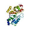
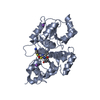
| Title | STRUCTURE OF BOVINE LIVER RHODANESE. I. STRUCTURE DETERMINATION AT 2.5 ANGSTROMS RESOLUTION AND A COMPARISON OF THE CONFORMATION AND SEQUENCE OF ITS TWO DOMAINS | ||||||
 Components Components | RHODANESE | ||||||
 Keywords Keywords | TRANSFERASE(THIOSULFATE / CYANIDE SULFUR) | ||||||
| Function / homology |  Function and homology information Function and homology informationrRNA transport / 3-mercaptopyruvate sulfurtransferase activity / thiosulfate sulfurtransferase / thiosulfate sulfurtransferase activity / rRNA import into mitochondrion / 5S rRNA binding / mitochondrial matrix / mitochondrionSimilarity search - Function | ||||||
| Biological species |  Bos taurus (cattle) Bos taurus (cattle) | ||||||
| Method | X-RAY DIFFRACTION / Resolution: 2.5 Å | ||||||
 Authors Authors | Hol, W.G.J. / Ploegman, J.H. / Kalk, K.H. / Drent, G. | ||||||
 Citation Citation | Journal: J.Mol.Biol. / Year: 1978 Title: Structure of bovine liver rhodanese. I. Structure determination at 2.5 A resolution and a comparison of the conformation and sequence of its two domains. Authors: Ploegman, J.H. / Drent, G. / Kalk, K.H. / Hol, W.G. #1: Journal: Biochemistry / Year: 1983Title: Binding of Metal Cyanide Complexes to Bovine Liver Rhodanese in the Crystalline State Authors: Lijk, L.J. / Kalk, K.H. / Brandenburg, N.P. / Hol, W.G.J. #2: Journal: J.Mol.Biol. / Year: 1979Title: The Structure of Bovine Liver Rhodanese. II. The Active Site in the Sulfur-Substituted and the Sulfur-Free Enzyme Authors: Ploegman, J.H. / Drent, G. / Kalk, K.H. / Hol, W.G.J. #3: Journal: J.Mol.Biol. / Year: 1975Title: The Double Domain Structure of Rhodanese Authors: Bergsma, J. / Hol, W.G.J. / Jansonius, J.N. / Kalk, K.H. / Ploegman, J.H. / Smit, J.D.G. #4: Journal: Thesis / Year: 1977Title: The Three-Dimensional Structure of Bovine Liver Rhodanese Authors: Ploegman, J.H. #5: Journal: Isr.J.Chem. / Year: 1974Title: The Structure of Rhodanese at 4 Angstroms Resolution. The Conformation of the Polypeptide Chain Authors: Smit, J.D.G. / Ploegman, J.H. / Pierrot, M. / Kalk, K.H. / Jansonius, J.N. / Drenth, J. #7: Journal: Biochem.Biophys.Res.Commun. / Year: 1971Title: Crystallographic Data for Rhodanese from Bovine Liver Authors: Drenth, J. / Smit, J.D.G. | ||||||
| History |
|
- Structure visualization
| Structure viewer | Molecule: MolmilJmol/JSmol |
|---|
- Downloads & links
-Download
| PDBx/mmCIF format | 1rhd.cif.gz | 67.2 KB | Display | PDBx/mmCIF format |
|---|---|---|---|---|
| PDB format | pdb1rhd.ent.gz | 46.6 KB | Display | PDB format |
| PDBx/mmJSON format | 1rhd.json.gz | Tree view | PDBx/mmJSON format | |
| Others |  Other downloads Other downloads |
-Validation report
| Arichive directory | https://data.pdbj.org/pub/pdb/validation_reports/rh/1rhdftp://data.pdbj.org/pub/pdb/validation_reports/rh/1rhd | HTTPS FTP |
|---|
-Related structure data
| Similar structure data |
|---|


-Links
 PDBj
PDBj- Assembly
| Deposited unit | 
| ||||||||
|---|---|---|---|---|---|---|---|---|---|
| 1 |
| ||||||||
| Unit cell |
|
-Components
| #1: Protein | Mass: 32982.375 Da / Num. of mol.: 1 Source method: isolated from a genetically manipulated source Source: (gene. exp.) Bos taurus (cattle) / References: UniProt: P00586, thiosulfate sulfurtransferase |
|---|
-Experimental details
-Experiment
| Experiment | Method: X-RAY DIFFRACTION |
|---|
- Sample preparation
| Crystal | Density Matthews: 2.42 Å3/Da / Density % sol: 49.18 % | |||||||||||||||
|---|---|---|---|---|---|---|---|---|---|---|---|---|---|---|---|---|
| Crystal grow | *PLUS pH: 7.3 / Method: unknown | |||||||||||||||
| Components of the solutions | *PLUS
|
-Data collection
| Radiation | Scattering type: x-ray |
|---|---|
| Radiation wavelength | Relative weight: 1 |
- Processing
| Refinement | Highest resolution: 2.5 Å | ||||||||||||
|---|---|---|---|---|---|---|---|---|---|---|---|---|---|
| Refinement step | Cycle: LAST / Highest resolution: 2.5 Å
| ||||||||||||
| Refinement | *PLUS Num. reflection obs: 10102 | ||||||||||||
| Solvent computation | *PLUS | ||||||||||||
| Displacement parameters | *PLUS |
