[English] 日本語
Yorodumi
- PDB-6w4m: CRYSTAL STRUCTURE OF THE ADCC-POTENT, WEAKLY NEUTRALIZING HIV ENV... -

+
Open data


ID or keywords:

Loading...

-
Basic information

Entry
Database: PDB / ID: 6w4m
Title
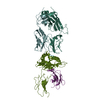
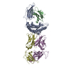
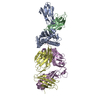
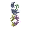
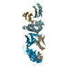
CRYSTAL STRUCTURE OF THE ADCC-POTENT, WEAKLY NEUTRALIZING HIV ENV CO-RECEPTOR BINDING SITE ANTIBODY N12-I2 FAB IN COMPLEX WITH HIV-1 CLADE A/E GP120 AND M48U1
Components
  • ANTI-HIV ANTIBODY N12-I2 FAB HEAVY CHAIN
  • ANTI-HIV ANTIBODY N12-I2 FAB LIGHT CHAIN
  • M48U1 CD4 MIMETIC PEPTIDE
  • clade A/E 93TH057 HIV-1 gp120 core
KeywordsVIRAL PROTEIN/IMMUNE SYSTEM / ADCC AND NEUTRALIZING ANTI-HIV-1 ENV ANTIBODY N12-I2 / CD4I ANTIBODY / FAB FRAGMENT / HIV-1 GP120 / IMMUNE SYSTEM / CLADE A/E 93TH057 / VIRAL PROTEIN-IMMUNE SYSTEM COMPLEX
Function / homologyGp120 core superfamily / Envelope glycoprotein GP120 / Human immunodeficiency virus 1, envelope glycoprotein Gp120 / viral envelope / clade A/E 93TH057 HIV-1 gp120 core
Function and homology information
Biological speciesHuman immunodeficiency virus 1
Homo sapiens (human)
synthetic construct (others)
MethodX-RAY DIFFRACTION / SYNCHROTRON / MOLECULAR REPLACEMENT / Resolution: 3.2 Å
AuthorsTolbert, W.D. / Gohain, N. / Pazgier, M.
Funding support United States, 3items
OrganizationGrant numberCountry
National Institutes of Health/National Institute Of Allergy and Infectious Diseases (NIH/NIAID)R01AI116274 United States
National Institutes of Health/National Institute Of Allergy and Infectious Diseases (NIH/NIAID)R01AI129769 United States
National Institutes of Health/National Institute Of Allergy and Infectious Diseases (NIH/NIAID)P01AI120756 United States
CitationJournal: Bmc Biol. / Year: 2020
Title: Defining rules governing recognition and Fc-mediated effector functions to the HIV-1 co-receptor binding site.
Authors: Tolbert, W.D. / Sherburn, R. / Gohain, N. / Ding, S. / Flinko, R. / Orlandi, C. / Ray, K. / Finzi, A. / Lewis, G.K. / Pazgier, M.
History
DepositionMar 11, 2020Deposition site: RCSB / Processing site: RCSB
SupersessionAug 5, 2020ID: 5UWE
Revision 1.0Aug 5, 2020Provider: repository / Type: Initial release
Revision 1.1Oct 18, 2023Group: Data collection / Database references / Refinement description
Category: chem_comp_atom / chem_comp_bond ...chem_comp_atom / chem_comp_bond / database_2 / pdbx_initial_refinement_model
Item: _database_2.pdbx_DOI / _database_2.pdbx_database_accession
Revision 1.2Nov 15, 2023Group: Data collection / Category: chem_comp_atom / chem_comp_bond / Item: _chem_comp_atom.atom_id / _chem_comp_bond.atom_id_2

-
Structure visualization

Structure viewerMolecule:
MolmilJmol/JSmol

Downloads & links

-
Assembly

Deposited unit
G: clade A/E 93TH057 HIV-1 gp120 core
L: ANTI-HIV ANTIBODY N12-I2 FAB LIGHT CHAIN
H: ANTI-HIV ANTIBODY N12-I2 FAB HEAVY CHAIN
N: M48U1 CD4 MIMETIC PEPTIDE
hetero molecules


Theoretical massNumber of molelcules
Total (without water)93,45713
Polymers91,4664
Non-polymers1,9919
Water0
1


  • Idetical with deposited unit
  • defined by author&software
  • Evidence: gel filtration
TypeNameSymmetry operationNumber
identity operation1_555x,y,z1
Buried area6530 Å2
ΔGint-37 kcal/mol
Surface area35810 Å2
MethodPISA
Unit cell
Length a, b, c (Å)52.687, 69.522, 213.468
Angle α, β, γ (deg.)90.00, 90.00, 90.00
Int Tables number19
Space group name H-MP212121

-
Components

#1: Protein clade A/E 93TH057 HIV-1 gp120 core


Mass: 39160.367 Da / Num. of mol.: 1 / Mutation: H375S
Source method: isolated from a genetically manipulated source
Source: (gene. exp.) Human immunodeficiency virus 1 / Gene: HIV-1 Env / Cell (production host): HEK 293 GnT1- / Production host: Homo sapiens (human) / References: UniProt: A0A0M3KKW9
#2: Antibody ANTI-HIV ANTIBODY N12-I2 FAB LIGHT CHAIN


Mass: 23437.988 Da / Num. of mol.: 1
Source method: isolated from a genetically manipulated source
Source: (gene. exp.) Homo sapiens (human) / Cell (production host): HEK 293 / Production host: Homo sapiens (human)
#3: Antibody ANTI-HIV ANTIBODY N12-I2 FAB HEAVY CHAIN


Mass: 25810.697 Da / Num. of mol.: 1
Source method: isolated from a genetically manipulated source
Source: (gene. exp.) Homo sapiens (human) / Cell (production host): HEK 293 / Production host: Homo sapiens (human)
#4: Protein/peptide M48U1 CD4 MIMETIC PEPTIDE


Mass: 3056.804 Da / Num. of mol.: 1 / Source method: obtained synthetically / Source: (synth.) synthetic construct (others)
#5: Sugar
ChemComp-NAG / 2-acetamido-2-deoxy-beta-D-glucopyranose / N-acetyl-beta-D-glucosamine / 2-acetamido-2-deoxy-beta-D-glucose / 2-acetamido-2-deoxy-D-glucose / 2-acetamido-2-deoxy-glucose / N-ACETYL-D-GLUCOSAMINE / N-Acetylglucosamine


Type: D-saccharide, beta linking / Mass: 221.208 Da / Num. of mol.: 9
Source method: isolated from a genetically manipulated source
Formula: C8H15NO6
IdentifierTypeProgram
DGlcpNAcbCONDENSED IUPAC CARBOHYDRATE SYMBOLGMML 1.0
N-acetyl-b-D-glucopyranosamineCOMMON NAMEGMML 1.0
b-D-GlcpNAcIUPAC CARBOHYDRATE SYMBOLPDB-CARE 1.0
GlcNAcSNFG CARBOHYDRATE SYMBOLGMML 1.0
Has ligand of interestY

-
Experimental details

-
Experiment

ExperimentMethod: X-RAY DIFFRACTION / Number of used crystals: 1

-
Sample preparation

CrystalDensity Matthews: 2.14 Å3/Da / Density % sol: 42.45 %
Crystal growTemperature: 294 K / Method: vapor diffusion, hanging drop / pH: 6.5 / Details: 15% PEG 6000 5% MPD 0.1 M MES pH 6.5

-
Data collection

Diffraction
IDMean temperature (K)Crystal-IDSerial crystal experiment
11001N
21001N
Diffraction source
SourceSiteBeamlineIDWavelength (Å)
SYNCHROTRONSSRL BL12-210.97946
SYNCHROTRONSSRL BL14-120.97946
Detector
TypeIDDetectorDate
DECTRIS PILATUS 6M1PIXELJan 7, 2016
MARMOSAIC 325 mm CCD2CCDJan 6, 2016
Radiation
IDMonochromatorProtocolMonochromatic (M) / Laue (L)Scattering typeWavelength-ID
1SI (111)SINGLE WAVELENGTHMx-ray1
2SI (111)SINGLE WAVELENGTHMx-ray2
Radiation wavelength
IDWavelength (Å)Relative weight
10.979461
21
ReflectionResolution: 3.2→50 Å / Num. obs: 12129 / % possible obs: 88.5 % / Redundancy: 4.3 % / CC1/2: 0.931 / Rmerge(I) obs: 0.321 / Rpim(I) all: 0.156 / Net I/σ(I): 6.3
Reflection shellResolution: 3.2→3.26 Å / Rmerge(I) obs: 0.521 / Mean I/σ(I) obs: 1.1 / Num. unique obs: 456 / CC1/2: 0.611 / Rpim(I) all: 0.434 / % possible all: 66.9

-
Processing

Software
NameVersionClassification
PHENIX1.17.1_3660refinement
REFMAC5.7.0032refinement
HKL-2000data reduction
HKL-2000data scaling
PHASERphasing
RefinementMethod to determine structure: MOLECULAR REPLACEMENT
Starting model: 5UWE

5uwe
PDB Unreleased entry


Resolution: 3.2→42.34 Å / SU ML: 0.42 / Cross valid method: FREE R-VALUE / σ(F): 1.35 / Phase error: 30.65
RfactorNum. reflection% reflection
Rfree0.3011 612 5.06 %
Rwork0.241 --
obs0.2439 12090 88.49 %
Solvent computationShrinkage radii: 0.9 Å / VDW probe radii: 1.11 Å
Refinement stepCycle: LAST / Resolution: 3.2→42.34 Å
ProteinNucleic acidLigandSolventTotal
Num. atoms6194 0 126 0 6320
Refine LS restraints
Refine-IDTypeDev idealNumber
X-RAY DIFFRACTIONf_bond_d0.0036452
X-RAY DIFFRACTIONf_angle_d0.6868759
X-RAY DIFFRACTIONf_dihedral_angle_d16.199924
X-RAY DIFFRACTIONf_chiral_restr0.0471013
X-RAY DIFFRACTIONf_plane_restr0.0051112
LS refinement shell
Resolution (Å)Rfactor RfreeNum. reflection RfreeRfactor RworkNum. reflection RworkRefine-ID% reflection obs (%)
3.2-3.520.31641260.26722055X-RAY DIFFRACTION65
3.52-4.030.31421650.27453058X-RAY DIFFRACTION96
4.03-5.070.28431560.22753137X-RAY DIFFRACTION97
5.07-42.340.30291650.23033228X-RAY DIFFRACTION95
Refinement TLS params.

Method: refined / Refine-ID: X-RAY DIFFRACTION

IDL112)L122)L132)L222)L232)L332)S11 (Å °)S12 (Å °)S13 (Å °)S21 (Å °)S22 (Å °)S23 (Å °)S31 (Å °)S32 (Å °)S33 (Å °)T112)T122)T132)T222)T232)T332)Origin x (Å)Origin y (Å)Origin z (Å)
12.6353-0.9643-0.97432.5678-0.74754.9917-0.2061-0.2032-0.23690.32850.19520.49270.1664-0.39880.09030.39030.0391-0.00120.39560.04040.6509-1.608723.9538-32.6751
23.16211.4032-1.71224.0718-0.79062.7636-0.46620.5654-0.414-1.04930.2176-0.5350.5201-0.07650.14930.7248-0.02220.09720.5004-0.09690.639534.090118.8914-88.2093
32.55811.6545-2.00942.7433-2.32333.3596-0.0290.01080.2065-0.2410.13990.20140.00870.0664-0.09460.5865-0.0448-0.01750.4412-0.0690.560631.554332.0481-76.1671
40.06020.04780.1016-0.14270.0212-0.10240.2767-0.325-1.1054-0.56850.0214-1.02581.08391.4963-00.84650.1209-0.01821.0765-0.01270.998720.502720.4641-33.7932
Refinement TLS group
IDRefine-IDRefine TLS-IDSelection details
1X-RAY DIFFRACTION1(chain 'G' and resid 47 through 492)
2X-RAY DIFFRACTION2(chain 'L' and resid 1 through 212)
3X-RAY DIFFRACTION3(chain 'H' and resid 2 through 216)
4X-RAY DIFFRACTION4(chain 'N' and resid 1 through 27)

+
About Yorodumi

-
News

-
Feb 9, 2022. New format data for meta-information of EMDB entries

New format data for meta-information of EMDB entries

  • Version 3 of the EMDB header file is now the official format.
  • The previous official version 1.9 will be removed from the archive.

Related info.:EMDB header

External links:wwPDB to switch to version 3 of the EMDB data model

-
Aug 12, 2020. Covid-19 info

Covid-19 info

URL: https://pdbj.org/emnavi/covid19.php

New page: Covid-19 featured information page in EM Navigator.

Related info.:Covid-19 info / Mar 5, 2020. Novel coronavirus structure data

+
Mar 5, 2020. Novel coronavirus structure data

Novel coronavirus structure data

Related info.:Yorodumi Speices / Aug 12, 2020. Covid-19 info

External links:COVID-19 featured content - PDBj / Molecule of the Month (242):Coronavirus Proteases

+
Jan 31, 2019. EMDB accession codes are about to change! (news from PDBe EMDB page)

EMDB accession codes are about to change! (news from PDBe EMDB page)

  • The allocation of 4 digits for EMDB accession codes will soon come to an end. Whilst these codes will remain in use, new EMDB accession codes will include an additional digit and will expand incrementally as the available range of codes is exhausted. The current 4-digit format prefixed with “EMD-” (i.e. EMD-XXXX) will advance to a 5-digit format (i.e. EMD-XXXXX), and so on. It is currently estimated that the 4-digit codes will be depleted around Spring 2019, at which point the 5-digit format will come into force.
  • The EM Navigator/Yorodumi systems omit the EMD- prefix.

Related info.:Q: What is EMD? / ID/Accession-code notation in Yorodumi/EM Navigator

External links:EMDB Accession Codes are Changing Soon! / Contact to PDBj

+
Jul 12, 2017. Major update of PDB

Major update of PDB

  • wwPDB released updated PDB data conforming to the new PDBx/mmCIF dictionary.
  • This is a major update changing the version number from 4 to 5, and with Remediation, in which all the entries are updated.
  • In this update, many items about electron microscopy experimental information are reorganized (e.g. em_software).
  • Now, EM Navigator and Yorodumi are based on the updated data.

External links:wwPDB Remediation / Enriched Model Files Conforming to OneDep Data Standards Now Available in the PDB FTP Archive

-
Yorodumi

Thousand views of thousand structures

  • Yorodumi is a browser for structure data from EMDB, PDB, SASBDB, etc.
  • This page is also the successor to EM Navigator detail page, and also detail information page/front-end page for Omokage search.
  • The word "yorodu" (or yorozu) is an old Japanese word meaning "ten thousand". "mi" (miru) is to see.

Related info.:EMDB / PDB / SASBDB / Comparison of 3 databanks / Yorodumi Search / Aug 31, 2016. New EM Navigator & Yorodumi / Yorodumi Papers / Jmol/JSmol / Function and homology information / Changes in new EM Navigator and Yorodumi

Read more