 Movie
Movie Controller


+ Open data

- Basic information
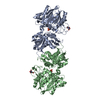
| Entry | Database: PDB / ID: 6c08 | ||||||
|---|---|---|---|---|---|---|---|
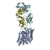
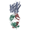
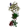
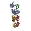
| Title | Zebrafish SLC38A9 with arginine bound in the cytosol open state | ||||||
 Components Components |
| ||||||
 Keywords Keywords |  MEMBRANE PROTEIN / transporter / conformational state / substrate binding / complex MEMBRANE PROTEIN / transporter / conformational state / substrate binding / complex | ||||||
| Function / homology |  Function and homology informationMacroautophagy / MTOR signalling / Energy dependent regulation of mTOR by LKB1-AMPK / TP53 Regulates Metabolic Genes / Regulation of PTEN gene transcription / Amino acids regulate mTORC1 / mTORC1-mediated signalling / asparagine transport / L-asparagine transmembrane transporter activity / sterol sensor activity ...Macroautophagy / MTOR signalling / Energy dependent regulation of mTOR by LKB1-AMPK / TP53 Regulates Metabolic Genes / Regulation of PTEN gene transcription / Amino acids regulate mTORC1 / mTORC1-mediated signalling / asparagine transport / L-asparagine transmembrane transporter activity / sterol sensor activity / amino acid sensor activity / L-arginine transmembrane transport / L-arginine transmembrane transporter activity / glutamine transport / L-glutamine transmembrane transporter activity / L-amino acid transmembrane transporter activity / L-leucine transmembrane transporter activity / amino acid transmembrane transport / L-leucine transport / amino acid transmembrane transporter activity / arginine binding / cholesterol binding / positive regulation of TOR signaling / positive regulation of TORC1 signaling / guanyl-nucleotide exchange factor activity / cellular response to amino acid stimulus / late endosome / late endosome membrane / lysosome / lysosomal membrane / metal ion binding Function and homology informationMacroautophagy / MTOR signalling / Energy dependent regulation of mTOR by LKB1-AMPK / TP53 Regulates Metabolic Genes / Regulation of PTEN gene transcription / Amino acids regulate mTORC1 / mTORC1-mediated signalling / asparagine transport / L-asparagine transmembrane transporter activity / sterol sensor activity ...Macroautophagy / MTOR signalling / Energy dependent regulation of mTOR by LKB1-AMPK / TP53 Regulates Metabolic Genes / Regulation of PTEN gene transcription / Amino acids regulate mTORC1 / mTORC1-mediated signalling / asparagine transport / L-asparagine transmembrane transporter activity / sterol sensor activity / amino acid sensor activity / L-arginine transmembrane transport / L-arginine transmembrane transporter activity / glutamine transport / L-glutamine transmembrane transporter activity / L-amino acid transmembrane transporter activity / L-leucine transmembrane transporter activity / amino acid transmembrane transport / L-leucine transport / amino acid transmembrane transporter activity / arginine binding / cholesterol binding / positive regulation of TOR signaling / positive regulation of TORC1 signaling / guanyl-nucleotide exchange factor activity / cellular response to amino acid stimulus / late endosome / late endosome membrane / lysosome / lysosomal membrane / metal ion bindingSimilarity search - Function | ||||||
| Biological species |  Danio rerio (zebrafish) Danio rerio (zebrafish) Mus musculus (house mouse) Mus musculus (house mouse) | ||||||
| Method | X-RAY DIFFRACTION / SYNCHROTRON / Resolution: 3.17 Å | ||||||
 Authors Authors | Lei, H.-T. / Gonen, T. | ||||||
 Citation Citation | Journal: Nat. Struct. Mol. Biol. / Year: 2018 Title: Crystal structure of arginine-bound lysosomal transporter SLC38A9 in the cytosol-open state. Authors: Lei, H.T. / Ma, J. / Sanchez Martinez, S. / Gonen, T. | ||||||
| History |
|
- Structure visualization
| Structure viewer | Molecule: MolmilJmol/JSmol |
|---|
- Downloads & links
-Download
| PDBx/mmCIF format | 6c08.cif.gz | 317.2 KB | Display | PDBx/mmCIF format |
|---|---|---|---|---|
| PDB format | pdb6c08.ent.gz | 259 KB | Display | PDB format |
| PDBx/mmJSON format | 6c08.json.gz | Tree view | PDBx/mmJSON format | |
| Others |  Other downloads Other downloads |
-Validation report
| Arichive directory | https://data.pdbj.org/pub/pdb/validation_reports/c0/6c08ftp://data.pdbj.org/pub/pdb/validation_reports/c0/6c08 | HTTPS FTP |
|---|
-Related structure data
| Similar structure data |
|---|


-Links
 PDBj
PDBj


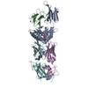
- Assembly
| Deposited unit | 
| ||||||||
|---|---|---|---|---|---|---|---|---|---|
| 1 | 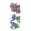
| ||||||||
| 2 | 
| ||||||||
| Unit cell |
|
-Components
| #1: Antibody | Mass: 22696.260 Da / Num. of mol.: 2 / Source method: isolated from a natural source Details: experimental sequence of heavy chains from antibody fragment is not determined because it is not discussed in the publication. Source: (natural) Mus musculus (house mouse)#2: Antibody | Mass: 23410.609 Da / Num. of mol.: 2 / Source method: isolated from a natural source Details: Experimental sequence of light chains from antibody fragment is not determined because it is not discussed in the publication. Source: (natural) Mus musculus (house mouse)#3: Protein | Mass: 54116.090 Da / Num. of mol.: 2 Source method: isolated from a genetically manipulated source Details: membrane protein / Source: (gene. exp.) Danio rerio (zebrafish) / Gene: slc38a9, zgc:154088 / Production host:   Spodoptera frugiperda (fall armyworm) / References: UniProt: Q08BA4 Spodoptera frugiperda (fall armyworm) / References: UniProt: Q08BA4#4: Chemical | ChemComp-ARG / | Arginine  Type: L-peptide linking / Mass: 175.209 Da / Num. of mol.: 1 / Source method: obtained synthetically / Formula: C6H15N4O2 Type: L-peptide linking / Mass: 175.209 Da / Num. of mol.: 1 / Source method: obtained synthetically / Formula: C6H15N4O2Sequence details | experimental sequence of antibody fragment is not determined | |
|---|
-Experimental details
-Experiment
| Experiment | Method: X-RAY DIFFRACTION / Number of used crystals: 1 |
|---|
- Sample preparation
| Crystal | Density Matthews: 4.42 Å3/Da / Density % sol: 72.15 % |
|---|---|
| Crystal grow | Temperature: 277 K / Method: vapor diffusion, hanging drop Details: PEG400, N-(2-Acetamido)iminodiacetic acid pH 7.2, lithium sulfate |
-Data collection
| Diffraction | Mean temperature: 100 K | ||||||||||||||||||||||||||||||
|---|---|---|---|---|---|---|---|---|---|---|---|---|---|---|---|---|---|---|---|---|---|---|---|---|---|---|---|---|---|---|---|
| Diffraction source | Source: SYNCHROTRON / Site: APS  / Beamline: 24-ID-C / Wavelength: 0.979 Å / Beamline: 24-ID-C / Wavelength: 0.979 Å | ||||||||||||||||||||||||||||||
| Detector | Type: DECTRIS PILATUS 6M-F / Detector: PIXEL / Date: Mar 18, 2017 | ||||||||||||||||||||||||||||||
| Radiation | Protocol: SINGLE WAVELENGTH / Monochromatic (M) / Laue (L): M / Scattering type: x-ray | ||||||||||||||||||||||||||||||
| Radiation wavelength | Wavelength: 0.979 Å / Relative weight: 1 | ||||||||||||||||||||||||||||||
| Reflection | Resolution: 3.1→156.5 Å / Num. obs: 63753 / % possible obs: 99.7 % / Redundancy: 8.3 % / Biso Wilson estimate: 114.41 Å2 / CC1/2: 0.997 / Rmerge(I) obs: 0.118 / Rpim(I) all: 0.044 / Rrim(I) all: 0.127 / Net I/σ(I): 8.3 / Num. measured all: 529570 / Scaling rejects: 391 | ||||||||||||||||||||||||||||||
| Reflection shell | Diffraction-ID: 1
|
- Processing
| Software |
| ||||||||||||||||||||||||||||||||||||||||||||||||||||||||||||||||||||||||||||||||||||||||||||||||||
|---|---|---|---|---|---|---|---|---|---|---|---|---|---|---|---|---|---|---|---|---|---|---|---|---|---|---|---|---|---|---|---|---|---|---|---|---|---|---|---|---|---|---|---|---|---|---|---|---|---|---|---|---|---|---|---|---|---|---|---|---|---|---|---|---|---|---|---|---|---|---|---|---|---|---|---|---|---|---|---|---|---|---|---|---|---|---|---|---|---|---|---|---|---|---|---|---|---|---|---|
| Refinement | Resolution: 3.17→156.498 Å / SU ML: 0.51 / Cross valid method: THROUGHOUT / σ(F): 1.34 / Phase error: 32.51 / Stereochemistry target values: ML
| ||||||||||||||||||||||||||||||||||||||||||||||||||||||||||||||||||||||||||||||||||||||||||||||||||
| Solvent computation | Shrinkage radii: 0.9 Å / VDW probe radii: 1.11 Å / Solvent model: FLAT BULK SOLVENT MODEL | ||||||||||||||||||||||||||||||||||||||||||||||||||||||||||||||||||||||||||||||||||||||||||||||||||
| Displacement parameters | Biso max: 202.97 Å2 / Biso mean: 113.9931 Å2 / Biso min: 45.24 Å2 | ||||||||||||||||||||||||||||||||||||||||||||||||||||||||||||||||||||||||||||||||||||||||||||||||||
| Refinement step | Cycle: final / Resolution: 3.17→156.498 Å
| ||||||||||||||||||||||||||||||||||||||||||||||||||||||||||||||||||||||||||||||||||||||||||||||||||
| LS refinement shell | Refine-ID: X-RAY DIFFRACTION / Rfactor Rfree error: 0 / Total num. of bins used: 13
|
