 Movie
Movie Controller

[English] 日本語
 Yorodumi
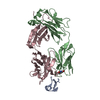
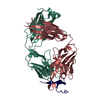
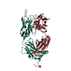
- PDB-6uvo: Structure of antibody 3G12 bound to the central conserved domain ... -
+ Open data

- Basic information
| Entry | Database: PDB / ID: 6uvo | ||||||
|---|---|---|---|---|---|---|---|
| Title | Structure of antibody 3G12 bound to the central conserved domain of RSV G | ||||||
 Components Components |
| ||||||
 Keywords Keywords |  VIRAL PROTEIN / IMMUNE SYSTEM / RSV / glycoprotein / G glycoprotein / viral attachment protein / antibody VIRAL PROTEIN / IMMUNE SYSTEM / RSV / glycoprotein / G glycoprotein / viral attachment protein / antibody | ||||||
| Function / homology |  Function and homology information Function and homology informationAssembly and release of respiratory syncytial virus (RSV) virions / Translation of respiratory syncytial virus mRNAs / Respiratory syncytial virus (RSV) attachment and entry / adhesion receptor-mediated virion attachment to host cell / RSV-host interactions / Maturation of hRSV A proteins / symbiont entry into host cell / virus-mediated perturbation of host defense response / host cell plasma membrane / virion membrane ...Assembly and release of respiratory syncytial virus (RSV) virions / Translation of respiratory syncytial virus mRNAs / Respiratory syncytial virus (RSV) attachment and entry / adhesion receptor-mediated virion attachment to host cell / RSV-host interactions / Maturation of hRSV A proteins / symbiont entry into host cell / virus-mediated perturbation of host defense response / host cell plasma membrane / virion membrane / extracellular region / plasma membraneSimilarity search - Function | ||||||
| Biological species |  Homo sapiens (human) Homo sapiens (human) Human respiratory syncytial virus A Human respiratory syncytial virus A | ||||||
| Method | X-RAY DIFFRACTION / SYNCHROTRON / MOLECULAR REPLACEMENT / Resolution: 2.9 Å | ||||||
 Authors Authors | Fedechkin, S.O. / George, N.L. / Nunez Castrejon, A.M. / Dillen, J. / Kauvar, L.M. / DuBois, R.M. | ||||||
| Funding support |  United States, 1items United States, 1items
| ||||||
 Citation Citation | Journal: J.Virol. / Year: 2020 Title: Conformational Flexibility in Respiratory Syncytial Virus G Neutralizing Epitopes. Authors: Fedechkin, S.O. / George, N.L. / Nunez Castrejon, A.M. / Dillen, J.R. / Kauvar, L.M. / DuBois, R.M. | ||||||
| History |
|
- Structure visualization
| Structure viewer | Molecule: MolmilJmol/JSmol |
|---|
- Downloads & links
-Download
| PDBx/mmCIF format | 6uvo.cif.gz | 194.3 KB | Display | PDBx/mmCIF format |
|---|---|---|---|---|
| PDB format | pdb6uvo.ent.gz | 155.1 KB | Display | PDB format |
| PDBx/mmJSON format | 6uvo.json.gz | Tree view | PDBx/mmJSON format | |
| Others |  Other downloads Other downloads |
-Validation report
| Arichive directory | https://data.pdbj.org/pub/pdb/validation_reports/uv/6uvoftp://data.pdbj.org/pub/pdb/validation_reports/uv/6uvo | HTTPS FTP |
|---|
-Related structure data
| Related structure data | 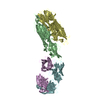 5k59S S: Starting model for refinement |
|---|---|
| Similar structure data |


-Links
 PDBj
PDBj

- Assembly
| Deposited unit | 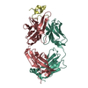
| ||||||||
|---|---|---|---|---|---|---|---|---|---|
| 1 |
| ||||||||
| Unit cell |
|
-Components
| #1: Antibody | Mass: 23218.771 Da / Num. of mol.: 1 Source method: isolated from a genetically manipulated source Source: (gene. exp.) Homo sapiens (human) / Production host:   Cricetulus griseus (Chinese hamster) Cricetulus griseus (Chinese hamster) |
|---|---|
| #2: Antibody | Mass: 24838.816 Da / Num. of mol.: 1 Source method: isolated from a genetically manipulated source Source: (gene. exp.) Homo sapiens (human) / Production host: Cricetulus griseus (Chinese hamster) |
| #3: Protein/peptide | Mass: 5735.633 Da / Num. of mol.: 1 Source method: isolated from a genetically manipulated source Source: (gene. exp.) Human respiratory syncytial virus A (strain A2)Strain: A2 / Production host:  Escherichia coli (E. coli) / References: UniProt: P03423 Escherichia coli (E. coli) / References: UniProt: P03423 |
-Experimental details
-Experiment
| Experiment | Method: X-RAY DIFFRACTION / Number of used crystals: 1 |
|---|
- Sample preparation
| Crystal | Density Matthews: 5.19 Å3/Da / Density % sol: 76.29 % |
|---|---|
| Crystal grow | Temperature: 277.15 K / Method: vapor diffusion / pH: 4.4 Details: 1.8M Ammonium Sulfate, 100mM sodium acetate trihydrate pH4.4 |
-Data collection
| Diffraction | Mean temperature: 100 K / Serial crystal experiment: N | |||||||||||||||||||||||||||
|---|---|---|---|---|---|---|---|---|---|---|---|---|---|---|---|---|---|---|---|---|---|---|---|---|---|---|---|---|
| Diffraction source | Source: SYNCHROTRON / Site: ALS / Beamline: 8.3.1 / Wavelength: 1.11503 Å | |||||||||||||||||||||||||||
| Detector | Type: DECTRIS PILATUS3 6M / Detector: PIXEL / Date: Feb 24, 2018 / Details: Pilatus3 S, 25Hz, S/N 60-0134 | |||||||||||||||||||||||||||
| Radiation | Monochromator: Si(111) Khozu / Protocol: SINGLE WAVELENGTH / Monochromatic (M) / Laue (L): M / Scattering type: x-ray | |||||||||||||||||||||||||||
| Radiation wavelength | Wavelength: 1.11503 Å / Relative weight: 1 | |||||||||||||||||||||||||||
| Reflection | Resolution: 2.9→74.531 Å / Num. obs: 23682 / % possible obs: 99.5 % / Redundancy: 3.9 % / Biso Wilson estimate: 66.24 Å2 / CC1/2: 0.993 / Rmerge(I) obs: 0.109 / Rpim(I) all: 0.062 / Rrim(I) all: 0.126 / Net I/σ(I): 9.4 | |||||||||||||||||||||||||||
| Reflection shell | Diffraction-ID: 1 / Redundancy: 3.8 %
|
- Processing
| Software |
| ||||||||||||||||||||||||||||||||||||||||||||||||||||||||||||||||||||||||||||||||||||||||||||||||||||
|---|---|---|---|---|---|---|---|---|---|---|---|---|---|---|---|---|---|---|---|---|---|---|---|---|---|---|---|---|---|---|---|---|---|---|---|---|---|---|---|---|---|---|---|---|---|---|---|---|---|---|---|---|---|---|---|---|---|---|---|---|---|---|---|---|---|---|---|---|---|---|---|---|---|---|---|---|---|---|---|---|---|---|---|---|---|---|---|---|---|---|---|---|---|---|---|---|---|---|---|---|---|
| Refinement | Method to determine structure: MOLECULAR REPLACEMENT Starting model: 5K59 Resolution: 2.9→74.53 Å / SU ML: 0.32 / Cross valid method: THROUGHOUT / σ(F): 1.33 / Phase error: 20.56
| ||||||||||||||||||||||||||||||||||||||||||||||||||||||||||||||||||||||||||||||||||||||||||||||||||||
| Solvent computation | Shrinkage radii: 0.9 Å / VDW probe radii: 1.11 Å | ||||||||||||||||||||||||||||||||||||||||||||||||||||||||||||||||||||||||||||||||||||||||||||||||||||
| Displacement parameters | Biso max: 190.21 Å2 / Biso mean: 63.3008 Å2 / Biso min: 27.71 Å2 | ||||||||||||||||||||||||||||||||||||||||||||||||||||||||||||||||||||||||||||||||||||||||||||||||||||
| Refinement step | Cycle: final / Resolution: 2.9→74.53 Å
| ||||||||||||||||||||||||||||||||||||||||||||||||||||||||||||||||||||||||||||||||||||||||||||||||||||
| Refine LS restraints |
| ||||||||||||||||||||||||||||||||||||||||||||||||||||||||||||||||||||||||||||||||||||||||||||||||||||
| LS refinement shell | Refine-ID: X-RAY DIFFRACTION / Rfactor Rfree error: 0
| ||||||||||||||||||||||||||||||||||||||||||||||||||||||||||||||||||||||||||||||||||||||||||||||||||||
| Refinement TLS params. | Method: refined / Refine-ID: X-RAY DIFFRACTION
| ||||||||||||||||||||||||||||||||||||||||||||||||||||||||||||||||||||||||||||||||||||||||||||||||||||
| Refinement TLS group |
|
