 Movie
Movie Controller

[English] 日本語
 Yorodumi
- PDB-2j5l: Structure of a Plasmodium falciparum apical membrane antigen 1-Fa... -
+ Open data

- Basic information
| Entry | Database: PDB / ID: 2j5l | |||||||||
|---|---|---|---|---|---|---|---|---|---|---|
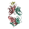
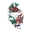
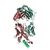
| Title | Structure of a Plasmodium falciparum apical membrane antigen 1-Fab F8. 12.19 complex | |||||||||
 Components Components |
| |||||||||
 Keywords Keywords |  IMMUNE SYSTEM / MALARIA VACCINE CANDIDATE / APICAL MEMBRANE ANTIGEN 1 / HYPOTHETICAL PROTEIN / IMMUNOGLOBULIN DOMAIN / MEMBRANE / MEROZOITE / TRANSMEMBRANE / IMMUNOGLOBULIN C REGION / ANTIBODY CROSS-REACTIVITY IMMUNE SYSTEM / MALARIA VACCINE CANDIDATE / APICAL MEMBRANE ANTIGEN 1 / HYPOTHETICAL PROTEIN / IMMUNOGLOBULIN DOMAIN / MEMBRANE / MEROZOITE / TRANSMEMBRANE / IMMUNOGLOBULIN C REGION / ANTIBODY CROSS-REACTIVITY | |||||||||
| Function / homology |  Function and homology information Function and homology information | |||||||||
| Biological species |  PLASMODIUM FALCIPARUM (malaria parasite P. falciparum) PLASMODIUM FALCIPARUM (malaria parasite P. falciparum) MUS MUSCULUS (house mouse) MUS MUSCULUS (house mouse) | |||||||||
| Method | X-RAY DIFFRACTION / SYNCHROTRON / MOLECULAR REPLACEMENT / Resolution: 2.9 Å | |||||||||
 Authors Authors | Igonet, S. / Vulliez-Le Normand, B. / Faure, G. / Riottot, M.M. / Kocken, C.H.M. / Thomas, A.W. / Bentley, G.A. | |||||||||
 Citation Citation | Journal: J.Mol.Biol. / Year: 2007 Title: Cross-Reactivity Studies of an Anti-Plasmodium Vivax Apical Membrane Antigen 1 Monoclonal Antibody: Binding and Structural Characterisation. Authors: Igonet, S. / Vulliez-Le Normand, B. / Faure, G. / Riottot, M.M. / Kocken, C.H.M. / Thomas, A.W. / Bentley, G.A. | |||||||||
| History |
| |||||||||
| Remark 700 | SHEET THE SHEET STRUCTURE OF THIS MOLECULE IS BIFURCATED. IN ORDER TO REPRESENT THIS FEATURE IN ... SHEET THE SHEET STRUCTURE OF THIS MOLECULE IS BIFURCATED. IN ORDER TO REPRESENT THIS FEATURE IN THE SHEET RECORDS BELOW, TWO SHEETS ARE DEFINED. |
- Structure visualization
| Structure viewer | Molecule: MolmilJmol/JSmol |
|---|
- Downloads & links
-Download
| PDBx/mmCIF format | 2j5l.cif.gz | 117.1 KB | Display | PDBx/mmCIF format |
|---|---|---|---|---|
| PDB format | pdb2j5l.ent.gz | 83.8 KB | Display | PDB format |
| PDBx/mmJSON format | 2j5l.json.gz | Tree view | PDBx/mmJSON format | |
| Others |  Other downloads Other downloads |
-Validation report
| Arichive directory | https://data.pdbj.org/pub/pdb/validation_reports/j5/2j5lftp://data.pdbj.org/pub/pdb/validation_reports/j5/2j5l | HTTPS FTP |
|---|
-Related structure data
| Related structure data |  2j4wSC S: Starting model for refinement C: citing same article ( |
|---|---|
| Similar structure data |


-Links
 PDBj
PDBj

- Assembly
| Deposited unit | 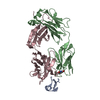
| ||||||||
|---|---|---|---|---|---|---|---|---|---|
| 1 |
| ||||||||
| Unit cell |
|
-Components
| #1: Protein | / PLASMODIUM FALCIPARUM APICAL MEMBRANE ANTIGEN 1 Mass: 67035.547 Da / Num. of mol.: 1 / Fragment: PFAMA1 ECTOPLASMIC REGION, RESIDUES 25-605 / Mutation: YES Source method: isolated from a genetically manipulated source Source: (gene. exp.) PLASMODIUM FALCIPARUM (malaria parasite P. falciparum)Strain: FVO / Plasmid: PPICZALPHAA / Production host:  PICHIA PASTORIS (fungus) / References: UniProt: Q9GVB7, UniProt: Q9BIM8*PLUS PICHIA PASTORIS (fungus) / References: UniProt: Q9GVB7, UniProt: Q9BIM8*PLUS |
|---|---|
| #2: Antibody | Mass: 23285.621 Da / Num. of mol.: 1 / Fragment: ANTIGEN-BINDING FRAGMENT FAB, LIGHT CHAIN / Source method: isolated from a natural source / Details: MONOCLONAL ANTIBODY ISOTYPE IS IGG1, KAPPA / Source: (natural) MUS MUSCULUS (house mouse) / Strain: BALB/C / References: UniProt: Q5XFY8*PLUS |
| #3: Antibody | Mass: 24189.033 Da / Num. of mol.: 1 / Fragment: ANTIGEN-BINDING FRAGMENT FAB, HEAVY CHAIN / Source method: isolated from a natural source / Details: MONOCLONAL ANTIBODY ISOTYPE IS IGG1, KAPPA. / Source: (natural) MUS MUSCULUS (house mouse) / Strain: BALB/C |
| Compound details | ENGINEERED RESIDUE IN CHAIN A, ASN 162 TO LYS ENGINEERED RESIDUE IN CHAIN A, THR 288 TO VAL ...ENGINEERED |
-Experimental details
-Experiment
| Experiment | Method: X-RAY DIFFRACTION / Number of used crystals: 1 |
|---|
- Sample preparation
| Crystal | Density Matthews: 2.2 Å3/Da / Density % sol: 45 % |
|---|---|
| Crystal grow | Temperature: 290 K / pH: 4.6 Details: CRYSTALLISATION TRIALS WERE CARRIED OUT WITH PFAMA1 ECTOPLASMIC CONSTRUCTION CORRESPONDING TO DOMAINS II-III. THE FAB FRAGMENT WAS INCUBATED IN SMALL STOICHIOMETRIC EXCESS WITH THE ...Details: CRYSTALLISATION TRIALS WERE CARRIED OUT WITH PFAMA1 ECTOPLASMIC CONSTRUCTION CORRESPONDING TO DOMAINS II-III. THE FAB FRAGMENT WAS INCUBATED IN SMALL STOICHIOMETRIC EXCESS WITH THE RECOMBINANT PROTEIN (1.2:1) BEFORE ADDING CRYSTALLISATION BUFFERS. CRYSTALLISATION DROPS WERE PREPARED BY MIXING 0.8 MICROL OF PROTEIN WITH 0.8 MICROL OF RESERVOIR BUFFER COMPRISING 12% PEG 6000 AND 0.1 M SODIUM ACETATE AT PH 4.6. THE FINAL PROTEIN CONCENTRATION WAS 3.2 MG/ML. CRYSTALS APPEARED AFTER 5 DAYS AT 17 DEGREE C. |
-Data collection
| Diffraction | Mean temperature: 100 K |
|---|---|
| Diffraction source | Source: SYNCHROTRON / Site: ESRF  / Beamline: ID14-2 / Wavelength: 0.949 / Beamline: ID14-2 / Wavelength: 0.949 |
| Detector | Type: ADSC CCD / Detector: CCD |
| Radiation | Protocol: SINGLE WAVELENGTH / Monochromatic (M) / Laue (L): M / Scattering type: x-ray |
| Radiation wavelength | Wavelength: 0.949 Å / Relative weight: 1 |
| Reflection | Resolution: 2.8→20 Å / Num. obs: 18102 / % possible obs: 96 % / Observed criterion σ(I): -3 / Redundancy: 21.2 % / Rmerge(I) obs: 0.25 / Net I/σ(I): 12.1 |
| Reflection shell | Resolution: 2.8→3 Å / Redundancy: 16.2 % / Rmerge(I) obs: 1.16 / Mean I/σ(I) obs: 2.7 / % possible all: 80.4 |
- Processing
| Software |
| ||||||||||||||||||||||||||||||||||||||||||||||||||||||||||||||||||||||||||||||||||||||||||||||||||||||||||||||||||||||||||||||||||||||||||||||||||||||||||||||||||||||||||||||||||||||
|---|---|---|---|---|---|---|---|---|---|---|---|---|---|---|---|---|---|---|---|---|---|---|---|---|---|---|---|---|---|---|---|---|---|---|---|---|---|---|---|---|---|---|---|---|---|---|---|---|---|---|---|---|---|---|---|---|---|---|---|---|---|---|---|---|---|---|---|---|---|---|---|---|---|---|---|---|---|---|---|---|---|---|---|---|---|---|---|---|---|---|---|---|---|---|---|---|---|---|---|---|---|---|---|---|---|---|---|---|---|---|---|---|---|---|---|---|---|---|---|---|---|---|---|---|---|---|---|---|---|---|---|---|---|---|---|---|---|---|---|---|---|---|---|---|---|---|---|---|---|---|---|---|---|---|---|---|---|---|---|---|---|---|---|---|---|---|---|---|---|---|---|---|---|---|---|---|---|---|---|---|---|---|---|
| Refinement | Method to determine structure: MOLECULAR REPLACEMENT Starting model: PDB ENTRY 2J4W Resolution: 2.9→20 Å / Cor.coef. Fo:Fc: 0.931 / Cor.coef. Fo:Fc free: 0.879 / SU B: 17.716 / SU ML: 0.328 / Cross valid method: THROUGHOUT / ESU R: 0.948 / ESU R Free: 0.39 / Stereochemistry target values: MAXIMUM LIKELIHOOD Details: HYDROGENS HAVE BEEN ADDED IN THE RIDING POSITIONS. FAB LIGHT AND HEAVY CHAIN ARE NUMBERED ACCORDING TO THE KABAT CONVENTION. ANTIGENS RESIDUES A303 TO A477 AND A513 TO A545 ARE DISORDERED IN ...Details: HYDROGENS HAVE BEEN ADDED IN THE RIDING POSITIONS. FAB LIGHT AND HEAVY CHAIN ARE NUMBERED ACCORDING TO THE KABAT CONVENTION. ANTIGENS RESIDUES A303 TO A477 AND A513 TO A545 ARE DISORDERED IN THE CRYSTAL STRUCTURE
| ||||||||||||||||||||||||||||||||||||||||||||||||||||||||||||||||||||||||||||||||||||||||||||||||||||||||||||||||||||||||||||||||||||||||||||||||||||||||||||||||||||||||||||||||||||||
| Solvent computation | Ion probe radii: 0.8 Å / Shrinkage radii: 0.8 Å / VDW probe radii: 1.2 Å / Solvent model: MASK | ||||||||||||||||||||||||||||||||||||||||||||||||||||||||||||||||||||||||||||||||||||||||||||||||||||||||||||||||||||||||||||||||||||||||||||||||||||||||||||||||||||||||||||||||||||||
| Displacement parameters | Biso mean: 61.04 Å2
| ||||||||||||||||||||||||||||||||||||||||||||||||||||||||||||||||||||||||||||||||||||||||||||||||||||||||||||||||||||||||||||||||||||||||||||||||||||||||||||||||||||||||||||||||||||||
| Refinement step | Cycle: LAST / Resolution: 2.9→20 Å
| ||||||||||||||||||||||||||||||||||||||||||||||||||||||||||||||||||||||||||||||||||||||||||||||||||||||||||||||||||||||||||||||||||||||||||||||||||||||||||||||||||||||||||||||||||||||
| Refine LS restraints |
|