 Movie
Movie Controller

[English] 日本語
 Yorodumi
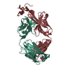
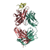
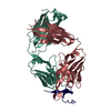
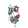
- PDB-1n64: Crystal structure analysis of the immunodominant antigenic site o... -
+ Open data

- Basic information
| Entry | Database: PDB / ID: 1n64 | ||||||
|---|---|---|---|---|---|---|---|
| Title | Crystal structure analysis of the immunodominant antigenic site on Hepatitis C virus protein bound to mAb 19D9D6 | ||||||
 Components Components |
| ||||||
 Keywords Keywords |  IMMUNE SYSTEM / ANTIBODY PEPTIDE COMPLEX IMMUNE SYSTEM / ANTIBODY PEPTIDE COMPLEX | ||||||
| Function / homology |  Function and homology informationhepacivirin / host cell mitochondrial membrane / host cell lipid droplet / symbiont-mediated suppression of host TRAF-mediated signal transduction / transformation of host cell by virus / symbiont-mediated perturbation of host cell cycle G1/S transition checkpoint / symbiont-mediated suppression of host JAK-STAT cascade via inhibition of STAT1 activity / symbiont-mediated suppression of host cytoplasmic pattern recognition receptor signaling pathway via inhibition of MAVS activity / negative regulation of DNA-binding transcription factor activity / SH3 domain binding ...hepacivirin / host cell mitochondrial membrane / host cell lipid droplet / symbiont-mediated suppression of host TRAF-mediated signal transduction / transformation of host cell by virus / symbiont-mediated perturbation of host cell cycle G1/S transition checkpoint / symbiont-mediated suppression of host JAK-STAT cascade via inhibition of STAT1 activity / symbiont-mediated suppression of host cytoplasmic pattern recognition receptor signaling pathway via inhibition of MAVS activity / negative regulation of DNA-binding transcription factor activity / SH3 domain binding / nucleoside-triphosphate phosphatase / protein complex oligomerization / monoatomic ion channel activity / viral nucleocapsid / clathrin-dependent endocytosis of virus by host cell / Hydrolases; Acting on peptide bonds (peptidases); Cysteine endopeptidases / RNA helicase activity / host cell perinuclear region of cytoplasm / host cell endoplasmic reticulum membrane / symbiont-mediated suppression of host type I interferon-mediated signaling pathway / RNA helicase / ribonucleoprotein complex / induction by virus of host autophagy / RNA-directed RNA polymerase / virus-mediated perturbation of host defense response / viral RNA genome replication / cysteine-type endopeptidase activity / RNA-dependent RNA polymerase activity / serine-type endopeptidase activity / fusion of virus membrane with host endosome membrane / viral envelope / host cell nucleus / virion attachment to host cell / host cell plasma membrane / virion membrane / structural molecule activity / ATP hydrolysis activity / proteolysis / RNA binding / zinc ion binding / ATP binding / membrane Function and homology informationhepacivirin / host cell mitochondrial membrane / host cell lipid droplet / symbiont-mediated suppression of host TRAF-mediated signal transduction / transformation of host cell by virus / symbiont-mediated perturbation of host cell cycle G1/S transition checkpoint / symbiont-mediated suppression of host JAK-STAT cascade via inhibition of STAT1 activity / symbiont-mediated suppression of host cytoplasmic pattern recognition receptor signaling pathway via inhibition of MAVS activity / negative regulation of DNA-binding transcription factor activity / SH3 domain binding ...hepacivirin / host cell mitochondrial membrane / host cell lipid droplet / symbiont-mediated suppression of host TRAF-mediated signal transduction / transformation of host cell by virus / symbiont-mediated perturbation of host cell cycle G1/S transition checkpoint / symbiont-mediated suppression of host JAK-STAT cascade via inhibition of STAT1 activity / symbiont-mediated suppression of host cytoplasmic pattern recognition receptor signaling pathway via inhibition of MAVS activity / negative regulation of DNA-binding transcription factor activity / SH3 domain binding / nucleoside-triphosphate phosphatase / protein complex oligomerization / monoatomic ion channel activity / viral nucleocapsid / clathrin-dependent endocytosis of virus by host cell / Hydrolases; Acting on peptide bonds (peptidases); Cysteine endopeptidases / RNA helicase activity / host cell perinuclear region of cytoplasm / host cell endoplasmic reticulum membrane / symbiont-mediated suppression of host type I interferon-mediated signaling pathway / RNA helicase / ribonucleoprotein complex / induction by virus of host autophagy / RNA-directed RNA polymerase / virus-mediated perturbation of host defense response / viral RNA genome replication / cysteine-type endopeptidase activity / RNA-dependent RNA polymerase activity / serine-type endopeptidase activity / fusion of virus membrane with host endosome membrane / viral envelope / host cell nucleus / virion attachment to host cell / host cell plasma membrane / virion membrane / structural molecule activity / ATP hydrolysis activity / proteolysis / RNA binding / zinc ion binding / ATP binding / membraneSimilarity search - Function | ||||||
| Biological species |  Mus musculus (house mouse) Mus musculus (house mouse) | ||||||
| Method | X-RAY DIFFRACTION / SYNCHROTRON / MOLECULAR REPLACEMENT / Resolution: 2.34 Å | ||||||
 Authors Authors | Menez, R. / Bossus, M. / Muller, B. / Sibai, G. / Dalbon, P. / Ducancel, F. / Jolivet-Reynaud, C. / Stura, E. | ||||||
 Citation Citation | Journal: J.Immunol. / Year: 2003 Title: Crystal structure of a hydrophobic immunodominant antigenic site on hepatitis C virus core protein complexed to monoclonal antibody 19D9D6. Authors: Menez, R. / Bossus, M. / Muller, B.H. / Sibai, G. / Dalbon, P. / Ducancel, F. / Jolivet-Reynaud, C. / Stura, E.A. | ||||||
| History |
|
- Structure visualization
| Structure viewer | Molecule: MolmilJmol/JSmol |
|---|
- Downloads & links
-Download
| PDBx/mmCIF format | 1n64.cif.gz | 99.5 KB | Display | PDBx/mmCIF format |
|---|---|---|---|---|
| PDB format | pdb1n64.ent.gz | 79.3 KB | Display | PDB format |
| PDBx/mmJSON format | 1n64.json.gz | Tree view | PDBx/mmJSON format | |
| Others |  Other downloads Other downloads |
-Validation report
| Arichive directory | https://data.pdbj.org/pub/pdb/validation_reports/n6/1n64ftp://data.pdbj.org/pub/pdb/validation_reports/n6/1n64 | HTTPS FTP |
|---|
-Related structure data
| Related structure data |  1nlbC  1dbaS  1hilS  1ucbS S: Starting model for refinement C: citing same article ( |
|---|---|
| Similar structure data |


-Links
 PDBj
PDBj


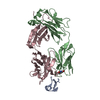
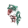
- Assembly
| Deposited unit | 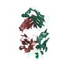
| ||||||||
|---|---|---|---|---|---|---|---|---|---|
| 1 |
| ||||||||
| Unit cell |
|
-Components
| #1: Antibody | Mass: 24334.051 Da / Num. of mol.: 1 / Source method: isolated from a natural source / Source: (natural) Mus musculus (house mouse) |
|---|---|
| #2: Antibody | Mass: 23563.430 Da / Num. of mol.: 1 Source method: isolated from a genetically manipulated source Source: (natural) Mus musculus (house mouse) |
| #3: Protein/peptide | Mass: 1640.929 Da / Num. of mol.: 1 / Source method: obtained synthetically / Details: SEQUENCE FROM HEPATITIS C VIRUS CORE PROTEIN References: UniProt: P29846, Hydrolases; Acting on peptide bonds (peptidases); Cysteine endopeptidases, hepacivirin, RNA-directed RNA polymerase |
| #4: Water | ChemComp-HOH / Water Mass: 18.015 Da / Num. of mol.: 202 / Source method: isolated from a natural source / Formula: H2O Mass: 18.015 Da / Num. of mol.: 202 / Source method: isolated from a natural source / Formula: H2O |
-Experimental details
-Experiment
| Experiment | Method: X-RAY DIFFRACTION / Number of used crystals: 1 |
|---|
- Sample preparation
| Crystal | Density Matthews: 2.36 Å3/Da / Density % sol: 47.89 % |
|---|---|
| Crystal grow | Temperature: 291 K / Method: vapor diffusion, sitting drop / pH: 9 Details: 10% MPEG 5000, 250mM NaCl, 50mM Tris-Hcl, pH 9.0, VAPOR DIFFUSION, SITTING DROP, temperature 291K |
| Crystal grow | *PLUS Method: unknown / Details: Stura, E.A., (2001) J. Crystal Growth, 232, 545. |
-Data collection
| Diffraction | Mean temperature: 100 K |
|---|---|
| Diffraction source | Source: SYNCHROTRON / Site: ESRF  / Beamline: BM30A / Wavelength: 1.009112 Å / Beamline: BM30A / Wavelength: 1.009112 Å |
| Detector | Type: MARRESEARCH / Detector: CCD / Date: Mar 30, 2001 / Details: mirrors |
| Radiation | Monochromator: Sagitally Focused Si (111) / Protocol: SINGLE WAVELENGTH / Monochromatic (M) / Laue (L): M / Scattering type: x-ray |
| Radiation wavelength | Wavelength: 1.009112 Å / Relative weight: 1 |
| Reflection | Resolution: 2.34→20 Å / Num. all: 19408 / Num. obs: 19303 / % possible obs: 98.9 % / Observed criterion σ(F): 2 / Observed criterion σ(I): 0 / Redundancy: 3 % / Rmerge(I) obs: 0.071 / Rsym value: 0.067 / Net I/σ(I): 9.2 |
| Reflection shell | Resolution: 2.34→2.39 Å / Redundancy: 1.7 % / Mean I/σ(I) obs: 1.5 / Num. unique all: 1179 / Rsym value: 0.233 / % possible all: 91.3 |
| Reflection | *PLUS Lowest resolution: 20 Å / Num. obs: 14779 / % possible obs: 98.8 % / Num. measured all: 17893 / Rmerge(I) obs: 0.067 |
| Reflection shell | *PLUS % possible obs: 91.3 % / Rmerge(I) obs: 0.233 |
- Processing
| Software |
| |||||||||||||||||||||||||
|---|---|---|---|---|---|---|---|---|---|---|---|---|---|---|---|---|---|---|---|---|---|---|---|---|---|---|
| Refinement | Method to determine structure: MOLECULAR REPLACEMENT Starting model: PDB ENTRIES 1UCB; 1DBA; 1HIL Resolution: 2.34→20 Å / Isotropic thermal model: ISOTROPIC / Cross valid method: THROUGHOUT / σ(F): 2 / Stereochemistry target values: Engh & Huber
| |||||||||||||||||||||||||
| Displacement parameters | Biso mean: 36.44 Å2 | |||||||||||||||||||||||||
| Refinement step | Cycle: LAST / Resolution: 2.34→20 Å
| |||||||||||||||||||||||||
| Refine LS restraints |
| |||||||||||||||||||||||||
| Refinement | *PLUS Lowest resolution: 20 Å / Rfactor Rwork: 0.19 | |||||||||||||||||||||||||
| Solvent computation | *PLUS | |||||||||||||||||||||||||
| Displacement parameters | *PLUS | |||||||||||||||||||||||||
| LS refinement shell | *PLUS Highest resolution: 2.34 Å / Lowest resolution: 2.39 Å / Rfactor Rfree: 0.268 / Rfactor Rwork: 0.216 |
