 Movie
Movie Controller


+ Open data

- Basic information
| Entry | Database: PDB / ID: 2hfg | ||||||
|---|---|---|---|---|---|---|---|
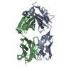
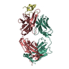
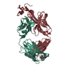
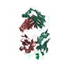
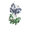
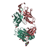
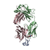
| Title | Crystal structure of hBR3 bound to CB3s-Fab | ||||||
 Components Components |
| ||||||
 Keywords Keywords |  IMMUNE SYSTEM / fab fragment / TNFRSF / antibody-receptor complex / CRD IMMUNE SYSTEM / fab fragment / TNFRSF / antibody-receptor complex / CRD | ||||||
| Function / homology |  Function and homology information Function and homology informationB cell costimulation / TNF receptor superfamily (TNFSF) members mediating non-canonical NF-kB pathway / positive regulation of T cell proliferation / positive regulation of B cell proliferation / tumor necrosis factor-mediated signaling pathway / T cell costimulation / TNFR2 non-canonical NF-kB pathway / signaling receptor activity / adaptive immune response / external side of plasma membrane ...B cell costimulation / TNF receptor superfamily (TNFSF) members mediating non-canonical NF-kB pathway / positive regulation of T cell proliferation / positive regulation of B cell proliferation / tumor necrosis factor-mediated signaling pathway / T cell costimulation / TNFR2 non-canonical NF-kB pathway / signaling receptor activity / adaptive immune response / external side of plasma membrane / membrane / plasma membraneSimilarity search - Function | ||||||
| Biological species |  Homo sapiens (human) Homo sapiens (human) | ||||||
| Method | X-RAY DIFFRACTION / SYNCHROTRON / MOLECULAR REPLACEMENT / Resolution: 2.61 Å | ||||||
 Authors Authors | Hymowitz, S.G. | ||||||
 Citation Citation | Journal: Blood / Year: 2006 Title: Synthetic anti-BR3 antibodies that mimic BAFF binding and target both human and murine B cells. Authors: Lee, C.V. / Hymowitz, S.G. / Wallweber, H.J. / Gordon, N.C. / Billeci, K.L. / Tsai, S.P. / Compaan, D.M. / Yin, J. / Gong, Q. / Kelley, R.F. / Deforge, L.E. / Martin, F. / Starovasnik, M.A. / Fuh, G. | ||||||
| History |
|
- Structure visualization
| Structure viewer | Molecule: MolmilJmol/JSmol |
|---|
- Downloads & links
-Download
| PDBx/mmCIF format | 2hfg.cif.gz | 99.8 KB | Display | PDBx/mmCIF format |
|---|---|---|---|---|
| PDB format | pdb2hfg.ent.gz | 77 KB | Display | PDB format |
| PDBx/mmJSON format | 2hfg.json.gz | Tree view | PDBx/mmJSON format | |
| Others |  Other downloads Other downloads |
-Validation report
| Arichive directory | https://data.pdbj.org/pub/pdb/validation_reports/hf/2hfgftp://data.pdbj.org/pub/pdb/validation_reports/hf/2hfg | HTTPS FTP |
|---|
-Related structure data


-Links
 PDBj
PDBj

- Assembly
| Deposited unit | 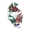
| ||||||||
|---|---|---|---|---|---|---|---|---|---|
| 1 |
| ||||||||
| Unit cell |
|
-Components
| #1: Antibody | Mass: 23250.775 Da / Num. of mol.: 1 Source method: isolated from a genetically manipulated source Source: (gene. exp.) Homo sapiens (human) / Description: PROTEIN SELECTED BY PHAGE DISPLAY / Cell line (production host): 4B8 / Production host:  Escherichia coli (E. coli) / References: UniProt: Q6PIH7 Escherichia coli (E. coli) / References: UniProt: Q6PIH7 |
|---|---|
| #2: Antibody | Mass: 24414.459 Da / Num. of mol.: 1 Source method: isolated from a genetically manipulated source Source: (gene. exp.) Homo sapiens (human) / Description: PROTEIN SELECTED BY PHAGE DISPLAY / Cell line (production host): 4B8 / Production host: Escherichia coli (E. coli) / References: UniProt: Q6N093 |
| #3: Protein | Mass: 5323.125 Da / Num. of mol.: 1 / Fragment: cysteine rich domain (residues 7-54) / Mutation: V20N, L27P Source method: isolated from a genetically manipulated source Source: (gene. exp.) Homo sapiens (human) / Gene: TNFRSF13C, BAFFR, BR3 / Plasmid: pET-32a / Production host: Escherichia coli (E. coli) / Strain (production host): Origami (DE3) / References: UniProt: Q96RJ3 |
| #4: Water | ChemComp-HOH / Water Mass: 18.015 Da / Num. of mol.: 27 / Source method: isolated from a natural source / Formula: H2O Mass: 18.015 Da / Num. of mol.: 27 / Source method: isolated from a natural source / Formula: H2O |
-Experimental details
-Experiment
| Experiment | Method: X-RAY DIFFRACTION / Number of used crystals: 1 |
|---|
- Sample preparation
| Crystal | Density Matthews: 2.47 Å3/Da / Density % sol: 50.12 % |
|---|---|
| Crystal grow | Details: Drops contained 2.1 microliters protein solution (pH 6.5) and 2.9 microliters of (0.1M citric acid pH 3.0, 24% PEG 3350, and 0.1 M Praseodymium (III) acetate) over a reservoir of 24% PEG ...Details: Drops contained 2.1 microliters protein solution (pH 6.5) and 2.9 microliters of (0.1M citric acid pH 3.0, 24% PEG 3350, and 0.1 M Praseodymium (III) acetate) over a reservoir of 24% PEG 3350. pH of final drop ~4.5. |
-Data collection
| Diffraction | Mean temperature: 100 K |
|---|---|
| Diffraction source | Source: SYNCHROTRON / Site: ALS  / Beamline: 5.0.1 / Wavelength: 1 / Beamline: 5.0.1 / Wavelength: 1 |
| Detector | Type: ADSC QUANTUM 315 / Detector: CCD / Date: Dec 17, 2004 |
| Radiation | Monochromator: Si(220) / Protocol: SINGLE WAVELENGTH / Monochromatic (M) / Laue (L): M / Scattering type: x-ray |
| Radiation wavelength | Wavelength: 1 Å / Relative weight: 1 |
| Reflection | Resolution: 2.6→50 Å / Num. all: 16168 / Num. obs: 16168 / % possible obs: 100 % / Observed criterion σ(F): 0 / Observed criterion σ(I): -3 / Rsym value: 0.064 / Net I/σ(I): 11.7 |
| Reflection shell | Resolution: 2.6→2.69 Å / Redundancy: 11.3 % / Mean I/σ(I) obs: 3.2 / Num. unique all: 1598 / Rsym value: 0.44 / % possible all: 100 |
- Processing
| Software |
| ||||||||||||||||||||||||||||||||||||||||||||||||||||||||||||||||||||||||||||||||||||||||||||||||||||||||||||||||||||||||||||||||||||||||||||||||||||||||||||||||||||||||||
|---|---|---|---|---|---|---|---|---|---|---|---|---|---|---|---|---|---|---|---|---|---|---|---|---|---|---|---|---|---|---|---|---|---|---|---|---|---|---|---|---|---|---|---|---|---|---|---|---|---|---|---|---|---|---|---|---|---|---|---|---|---|---|---|---|---|---|---|---|---|---|---|---|---|---|---|---|---|---|---|---|---|---|---|---|---|---|---|---|---|---|---|---|---|---|---|---|---|---|---|---|---|---|---|---|---|---|---|---|---|---|---|---|---|---|---|---|---|---|---|---|---|---|---|---|---|---|---|---|---|---|---|---|---|---|---|---|---|---|---|---|---|---|---|---|---|---|---|---|---|---|---|---|---|---|---|---|---|---|---|---|---|---|---|---|---|---|---|---|---|---|---|
| Refinement | Method to determine structure: MOLECULAR REPLACEMENT Starting model: CB2-Fab Resolution: 2.61→30 Å / Cor.coef. Fo:Fc: 0.945 / Cor.coef. Fo:Fc free: 0.914 / SU B: 23.633 / SU ML: 0.238 / TLS residual ADP flag: LIKELY RESIDUAL / Cross valid method: THROUGHOUT / σ(F): 0 / σ(I): -3 / ESU R: 1.455 / ESU R Free: 0.336 / Stereochemistry target values: MAXIMUM LIKELIHOOD / Details: HYDROGENS HAVE BEEN ADDED IN THE RIDING POSITIONS
| ||||||||||||||||||||||||||||||||||||||||||||||||||||||||||||||||||||||||||||||||||||||||||||||||||||||||||||||||||||||||||||||||||||||||||||||||||||||||||||||||||||||||||
| Solvent computation | Ion probe radii: 0.8 Å / Shrinkage radii: 0.8 Å / VDW probe radii: 1.4 Å / Solvent model: MASK | ||||||||||||||||||||||||||||||||||||||||||||||||||||||||||||||||||||||||||||||||||||||||||||||||||||||||||||||||||||||||||||||||||||||||||||||||||||||||||||||||||||||||||
| Displacement parameters | Biso mean: 47.436 Å2 | ||||||||||||||||||||||||||||||||||||||||||||||||||||||||||||||||||||||||||||||||||||||||||||||||||||||||||||||||||||||||||||||||||||||||||||||||||||||||||||||||||||||||||
| Refinement step | Cycle: LAST / Resolution: 2.61→30 Å
| ||||||||||||||||||||||||||||||||||||||||||||||||||||||||||||||||||||||||||||||||||||||||||||||||||||||||||||||||||||||||||||||||||||||||||||||||||||||||||||||||||||||||||
| Refine LS restraints |
| ||||||||||||||||||||||||||||||||||||||||||||||||||||||||||||||||||||||||||||||||||||||||||||||||||||||||||||||||||||||||||||||||||||||||||||||||||||||||||||||||||||||||||
| LS refinement shell | Resolution: 2.614→2.668 Å / Total num. of bins used: 25
| ||||||||||||||||||||||||||||||||||||||||||||||||||||||||||||||||||||||||||||||||||||||||||||||||||||||||||||||||||||||||||||||||||||||||||||||||||||||||||||||||||||||||||
| Refinement TLS params. | Method: refined / Refine-ID: X-RAY DIFFRACTION
| ||||||||||||||||||||||||||||||||||||||||||||||||||||||||||||||||||||||||||||||||||||||||||||||||||||||||||||||||||||||||||||||||||||||||||||||||||||||||||||||||||||||||||
| Refinement TLS group |
|
