 Movie
Movie Controller

[English] 日本語
 Yorodumi
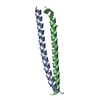
- PDB-6ucm: Transcription factor DeltaFosB bZIP domain self-assembly, type-II... -
+ Open data

- Basic information
| Entry | Database: PDB / ID: 6ucm | ||||||
|---|---|---|---|---|---|---|---|
| Title | Transcription factor DeltaFosB bZIP domain self-assembly, type-II crystal | ||||||
 Components Components | Protein fosB | ||||||
 Keywords Keywords |  TRANSCRIPTION / activator protein-1 / basic leucine zipper / bZIP / deltaFosB fos / jun / transcription factor / DNA-binding protein / coiled-coil TRANSCRIPTION / activator protein-1 / basic leucine zipper / bZIP / deltaFosB fos / jun / transcription factor / DNA-binding protein / coiled-coil | ||||||
| Function / homology |  Function and homology information Function and homology informationNGF-stimulated transcription / response to corticosterone / response to mechanical stimulus / cellular response to hormone stimulus / response to cAMP / cellular response to calcium ion / response to progesterone / female pregnancy / sequence-specific double-stranded DNA binding / DNA-binding transcription activator activity, RNA polymerase II-specific ...NGF-stimulated transcription / response to corticosterone / response to mechanical stimulus / cellular response to hormone stimulus / response to cAMP / cellular response to calcium ion / response to progesterone / female pregnancy / sequence-specific double-stranded DNA binding / DNA-binding transcription activator activity, RNA polymerase II-specific / Estrogen-dependent gene expression / transcription by RNA polymerase II / DNA-binding transcription factor activity, RNA polymerase II-specific / response to xenobiotic stimulus / RNA polymerase II cis-regulatory region sequence-specific DNA binding / intracellular membrane-bounded organelle / chromatin / regulation of transcription by RNA polymerase II / negative regulation of transcription by RNA polymerase II / DNA binding / nucleoplasm / nucleusSimilarity search - Function | ||||||
| Biological species |  Homo sapiens (human) Homo sapiens (human) | ||||||
| Method | X-RAY DIFFRACTION / SYNCHROTRON / MOLECULAR REPLACEMENT / Resolution: 2.424 Å | ||||||
 Authors Authors | Yin, Z. / Machius, M. / Rudenko, G. | ||||||
| Funding support |  United States, 1items United States, 1items
| ||||||
 Citation Citation | Journal: Curr Res Struct Biol / Year: 2020 Title: Self-assembly of the bZIP transcription factor Delta FosB. Authors: Yin, Z. / Venkannagari, H. / Lynch, H. / Aglyamova, G. / Bhandari, M. / Machius, M. / Nestler, E.J. / Robison, A.J. / Rudenko, G. | ||||||
| History |
|
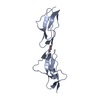
- Structure visualization
| Structure viewer | Molecule: MolmilJmol/JSmol |
|---|
- Downloads & links
-Download
| PDBx/mmCIF format | 6ucm.cif.gz | 107.2 KB | Display | PDBx/mmCIF format |
|---|---|---|---|---|
| PDB format | pdb6ucm.ent.gz | 84.2 KB | Display | PDB format |
| PDBx/mmJSON format | 6ucm.json.gz | Tree view | PDBx/mmJSON format | |
| Others |  Other downloads Other downloads |
-Validation report
| Arichive directory | https://data.pdbj.org/pub/pdb/validation_reports/uc/6ucmftp://data.pdbj.org/pub/pdb/validation_reports/uc/6ucm | HTTPS FTP |
|---|
-Related structure data
| Related structure data |  6uciC  6uclC  5vpeS C: citing same article ( S: Starting model for refinement |
|---|---|
| Similar structure data |


-Links
 PDBj
PDBj


- Assembly
| Deposited unit | 
| ||||||||
|---|---|---|---|---|---|---|---|---|---|
| 1 | 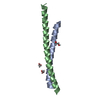
| ||||||||
| 2 | 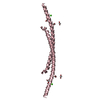
| ||||||||
| Unit cell |
|
-Components
| #1: Protein | Mass: 8267.290 Da / Num. of mol.: 3 Source method: isolated from a genetically manipulated source Details: alternative splice form; identical to the mouse sequence in the region cloned Source: (gene. exp.) Homo sapiens (human) / Gene: FOSB, G0S3 / Plasmid: pET21 NESG / Production host:  Escherichia coli (E. coli) / References: UniProt: P53539 Escherichia coli (E. coli) / References: UniProt: P53539#2: Chemical | ChemComp-EDO / Ethylene glycol  Mass: 62.068 Da / Num. of mol.: 6 / Source method: obtained synthetically / Formula: C2H6O2 Mass: 62.068 Da / Num. of mol.: 6 / Source method: obtained synthetically / Formula: C2H6O2#3: Chemical | ChemComp-CA / |   Mass: 40.078 Da / Num. of mol.: 1 / Source method: obtained synthetically / Formula: Ca Mass: 40.078 Da / Num. of mol.: 1 / Source method: obtained synthetically / Formula: Ca#4: Water | ChemComp-HOH / | Water Mass: 18.015 Da / Num. of mol.: 16 / Source method: isolated from a natural source / Formula: H2O Mass: 18.015 Da / Num. of mol.: 16 / Source method: isolated from a natural source / Formula: H2OHas ligand of interest | N | |
|---|
-Experimental details
-Experiment
| Experiment | Method: X-RAY DIFFRACTION / Number of used crystals: 1 |
|---|
- Sample preparation
| Crystal | Density Matthews: 3.1 Å3/Da / Density % sol: 60.38 % |
|---|---|
| Crystal grow | Temperature: 293 K / Method: vapor diffusion, hanging drop / pH: 7 / Details: 2 M ammonium sulfate, 5% (v/v) isopropanol |
-Data collection
| Diffraction | Mean temperature: 100 K / Serial crystal experiment: N | ||||||||||||||||||||||||||||||||||||||||||||||||||||||||||||||||||||||||||||||||||||||||||||||||||||||||||||||||||||||||||||||||||||||||||||||||||||||||||||||||||||||||
|---|---|---|---|---|---|---|---|---|---|---|---|---|---|---|---|---|---|---|---|---|---|---|---|---|---|---|---|---|---|---|---|---|---|---|---|---|---|---|---|---|---|---|---|---|---|---|---|---|---|---|---|---|---|---|---|---|---|---|---|---|---|---|---|---|---|---|---|---|---|---|---|---|---|---|---|---|---|---|---|---|---|---|---|---|---|---|---|---|---|---|---|---|---|---|---|---|---|---|---|---|---|---|---|---|---|---|---|---|---|---|---|---|---|---|---|---|---|---|---|---|---|---|---|---|---|---|---|---|---|---|---|---|---|---|---|---|---|---|---|---|---|---|---|---|---|---|---|---|---|---|---|---|---|---|---|---|---|---|---|---|---|---|---|---|---|---|---|---|---|
| Diffraction source | Source: SYNCHROTRON / Site: APS / Beamline: 19-ID / Wavelength: 0.987 Å | ||||||||||||||||||||||||||||||||||||||||||||||||||||||||||||||||||||||||||||||||||||||||||||||||||||||||||||||||||||||||||||||||||||||||||||||||||||||||||||||||||||||||
| Detector | Type: DECTRIS PILATUS3 6M / Detector: PIXEL / Date: Aug 19, 2015 | ||||||||||||||||||||||||||||||||||||||||||||||||||||||||||||||||||||||||||||||||||||||||||||||||||||||||||||||||||||||||||||||||||||||||||||||||||||||||||||||||||||||||
| Radiation | Protocol: SINGLE WAVELENGTH / Monochromatic (M) / Laue (L): M / Scattering type: x-ray | ||||||||||||||||||||||||||||||||||||||||||||||||||||||||||||||||||||||||||||||||||||||||||||||||||||||||||||||||||||||||||||||||||||||||||||||||||||||||||||||||||||||||
| Radiation wavelength | Wavelength: 0.987 Å / Relative weight: 1 | ||||||||||||||||||||||||||||||||||||||||||||||||||||||||||||||||||||||||||||||||||||||||||||||||||||||||||||||||||||||||||||||||||||||||||||||||||||||||||||||||||||||||
| Reflection | Resolution: 2.424→50 Å / Num. obs: 11940 / % possible obs: 100 % / Redundancy: 10.5 % / Biso Wilson estimate: 32.58 Å2 / Rmerge(I) obs: 0.086 / Rpim(I) all: 0.028 / Rrim(I) all: 0.091 / Χ2: 0.952 / Net I/σ(I): 9 | ||||||||||||||||||||||||||||||||||||||||||||||||||||||||||||||||||||||||||||||||||||||||||||||||||||||||||||||||||||||||||||||||||||||||||||||||||||||||||||||||||||||||
| Reflection shell | Diffraction-ID: 1 / % possible all: 100
|
- Processing
| Software |
| |||||||||||||||||||||||||||||||||||||||||||||||||||||||||||||||||||||||||||||||||||||||||||||||||||||||||||||||||||||||||||||
|---|---|---|---|---|---|---|---|---|---|---|---|---|---|---|---|---|---|---|---|---|---|---|---|---|---|---|---|---|---|---|---|---|---|---|---|---|---|---|---|---|---|---|---|---|---|---|---|---|---|---|---|---|---|---|---|---|---|---|---|---|---|---|---|---|---|---|---|---|---|---|---|---|---|---|---|---|---|---|---|---|---|---|---|---|---|---|---|---|---|---|---|---|---|---|---|---|---|---|---|---|---|---|---|---|---|---|---|---|---|---|---|---|---|---|---|---|---|---|---|---|---|---|---|---|---|---|
| Refinement | Method to determine structure: MOLECULAR REPLACEMENT Starting model: 5VPE Resolution: 2.424→44.384 Å / SU ML: 0.28 / Cross valid method: THROUGHOUT / σ(F): 1.35 / Phase error: 25.14
| |||||||||||||||||||||||||||||||||||||||||||||||||||||||||||||||||||||||||||||||||||||||||||||||||||||||||||||||||||||||||||||
| Solvent computation | Shrinkage radii: 0.9 Å / VDW probe radii: 1.11 Å | |||||||||||||||||||||||||||||||||||||||||||||||||||||||||||||||||||||||||||||||||||||||||||||||||||||||||||||||||||||||||||||
| Displacement parameters | Biso max: 142.22 Å2 / Biso mean: 57.5489 Å2 / Biso min: 14.86 Å2 | |||||||||||||||||||||||||||||||||||||||||||||||||||||||||||||||||||||||||||||||||||||||||||||||||||||||||||||||||||||||||||||
| Refinement step | Cycle: final / Resolution: 2.424→44.384 Å
| |||||||||||||||||||||||||||||||||||||||||||||||||||||||||||||||||||||||||||||||||||||||||||||||||||||||||||||||||||||||||||||
| Refine LS restraints |
| |||||||||||||||||||||||||||||||||||||||||||||||||||||||||||||||||||||||||||||||||||||||||||||||||||||||||||||||||||||||||||||
| LS refinement shell | Refine-ID: X-RAY DIFFRACTION / Rfactor Rfree error: 0
| |||||||||||||||||||||||||||||||||||||||||||||||||||||||||||||||||||||||||||||||||||||||||||||||||||||||||||||||||||||||||||||
| Refinement TLS params. | Method: refined / Refine-ID: X-RAY DIFFRACTION
| |||||||||||||||||||||||||||||||||||||||||||||||||||||||||||||||||||||||||||||||||||||||||||||||||||||||||||||||||||||||||||||
| Refinement TLS group |
|
