 Movie
Movie Controller

[English] 日本語
 Yorodumi
- PDB-3nz3: Crystal structure of the mucin-binding domain of Spr1345 from Str... -
+ Open data

- Basic information
| Entry | Database: PDB / ID: 3nz3 | ||||||
|---|---|---|---|---|---|---|---|
| Title | Crystal structure of the mucin-binding domain of Spr1345 from Streptococcus pneumoniae | ||||||
 Components Components | Putative uncharacterized protein | ||||||
 Keywords Keywords |  CELL ADHESION / All beta sheets / Ig-like fold / Mucin-binding / Mucin / Cell surface CELL ADHESION / All beta sheets / Ig-like fold / Mucin-binding / Mucin / Cell surface | ||||||
| Function / homology |  Function and homology information Function and homology information | ||||||
| Biological species |   Streptococcus pneumoniae (bacteria) Streptococcus pneumoniae (bacteria) | ||||||
| Method | X-RAY DIFFRACTION / SAD / Resolution: 2 Å | ||||||
 Authors Authors | Du, Y. / He, Y.-X. / Zhang, Z.-Y. / Yang, Y.-H. / Shi, W.-W. / Frolet, C. / Guilmi, A.M. / Vernet, T. / Zhou, C.-Z. / Chen, Y. | ||||||
 Citation Citation | Journal: J.Struct.Biol. / Year: 2011 Title: Crystal structure of the mucin-binding domain of Spr1345 from Streptococcus pneumoniae Authors: Du, Y. / He, Y.-X. / Zhang, Z.-Y. / Yang, Y.-H. / Shi, W.-W. / Frolet, C. / Guilmi, A.M. / Vernet, T. / Zhou, C.-Z. / Chen, Y. | ||||||
| History |
|
- Structure visualization
| Structure viewer | Molecule: MolmilJmol/JSmol |
|---|
- Downloads & links
-Download
| PDBx/mmCIF format | 3nz3.cif.gz | 35.6 KB | Display | PDBx/mmCIF format |
|---|---|---|---|---|
| PDB format | pdb3nz3.ent.gz | 24.7 KB | Display | PDB format |
| PDBx/mmJSON format | 3nz3.json.gz | Tree view | PDBx/mmJSON format | |
| Others |  Other downloads Other downloads |
-Validation report
| Arichive directory | https://data.pdbj.org/pub/pdb/validation_reports/nz/3nz3ftp://data.pdbj.org/pub/pdb/validation_reports/nz/3nz3 | HTTPS FTP |
|---|
-Related structure data
| Similar structure data |
|---|


-Links
 PDBj
PDBj- Assembly
| Deposited unit | 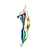
| ||||||||||||
|---|---|---|---|---|---|---|---|---|---|---|---|---|---|
| 1 |
| ||||||||||||
| Unit cell |
| ||||||||||||
| Components on special symmetry positions |
|
-Components
| #1: Protein | Mass: 12738.092 Da / Num. of mol.: 1 / Fragment: Mucin-binding domain Source method: isolated from a genetically manipulated source Source: (gene. exp.) Streptococcus pneumoniae (bacteria) / Strain: R6 / Gene: spr1345 / Plasmid: pET28a / Production host: Escherichia coli (E. coli) / Strain (production host): BL21-Ril / References: UniProt: Q8CYK3 |
|---|---|
| #2: Chemical | ChemComp-PGE / Polyethylene glycol  Mass: 150.173 Da / Num. of mol.: 1 / Source method: obtained synthetically / Formula: C6H14O4 Mass: 150.173 Da / Num. of mol.: 1 / Source method: obtained synthetically / Formula: C6H14O4 |
| #3: Chemical | ChemComp-SO4 / Sulfate  Mass: 96.063 Da / Num. of mol.: 1 / Source method: obtained synthetically / Formula: SO4 Mass: 96.063 Da / Num. of mol.: 1 / Source method: obtained synthetically / Formula: SO4 |
| #4: Water | ChemComp-HOH / Water Mass: 18.015 Da / Num. of mol.: 150 / Source method: isolated from a natural source / Formula: H2O Mass: 18.015 Da / Num. of mol.: 150 / Source method: isolated from a natural source / Formula: H2O |
-Experimental details
-Experiment
| Experiment | Method: X-RAY DIFFRACTION / Number of used crystals: 1 |
|---|
- Sample preparation
| Crystal | Density Matthews: 2.86 Å3/Da / Density % sol: 56.96 % |
|---|---|
| Crystal grow | Temperature: 289 K / Method: vapor diffusion, hanging drop / pH: 8.5 Details: 22% PEG 4000, 0.1M Tris-HCl, 0.2M Lithium Sulfate, pH 8.5, VAPOR DIFFUSION, HANGING DROP, temperature 289K |
-Data collection
| Diffraction | Mean temperature: 100 K |
|---|---|
| Diffraction source | Source: ROTATING ANODE / Type: RIGAKU MICROMAX-007 HF / Wavelength: 1.54178 Å |
| Detector | Type: MAR scanner 345 mm plate / Detector: IMAGE PLATE / Date: Oct 8, 2008 |
| Radiation | Protocol: SINGLE WAVELENGTH / Monochromatic (M) / Laue (L): M / Scattering type: x-ray |
| Radiation wavelength | Wavelength: 1.54178 Å / Relative weight: 1 |
| Reflection | Resolution: 2→37.48 Å / Num. obs: 10342 / % possible obs: 100 % / Observed criterion σ(F): 0 / Observed criterion σ(I): 0 / Redundancy: 8.4 % / Biso Wilson estimate: 26.8 Å2 / Rmerge(I) obs: 0.05 / Net I/σ(I): 29.2 |
| Reflection shell | Resolution: 2→2.11 Å / Redundancy: 8.3 % / Rmerge(I) obs: 0.05 / Mean I/σ(I) obs: 4.3 / Num. unique all: 1467 / % possible all: 100 |
- Processing
| Software |
| ||||||||||||||||||
|---|---|---|---|---|---|---|---|---|---|---|---|---|---|---|---|---|---|---|---|
| Refinement | Method to determine structure: SAD / Resolution: 2→25.78 Å / Isotropic thermal model: Isotropic / Cross valid method: THROUGHOUT / Stereochemistry target values: Engh & Huber
| ||||||||||||||||||
| Refinement step | Cycle: LAST / Resolution: 2→25.78 Å
|
