 Movie
Movie Controller

[English] 日本語
 Yorodumi
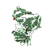
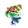
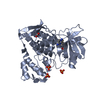
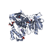
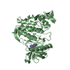
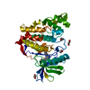
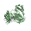
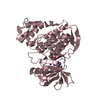
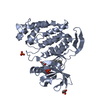
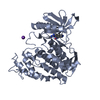
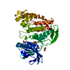
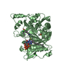
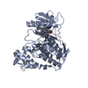
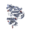
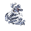
- PDB-6tw2: Re-refined crystal structure of di-phosphorylated human CLK1 in c... -
+ Open data

- Basic information
| Entry | Database: PDB / ID: 6tw2 | ||||||
|---|---|---|---|---|---|---|---|
| Title | Re-refined crystal structure of di-phosphorylated human CLK1 in complex with a novel substituted indole inhibitor | ||||||
 Components Components | Dual specificity protein kinase CLK1 | ||||||
 Keywords Keywords |  TRANSFERASE / SERINE/THREONINE-PROTEIN KINASE / TYROSINE-PROTEIN KINASE / NUCLEUS TRANSFERASE / SERINE/THREONINE-PROTEIN KINASE / TYROSINE-PROTEIN KINASE / NUCLEUS | ||||||
| Function / homology |  Function and homology informationdual-specificity kinase / regulation of RNA splicing / protein serine/threonine/tyrosine kinase activity / non-membrane spanning protein tyrosine kinase activity / protein tyrosine kinase activity / phosphorylation / protein serine kinase activity / protein serine/threonine kinase activity / ATP binding / nucleus Function and homology informationdual-specificity kinase / regulation of RNA splicing / protein serine/threonine/tyrosine kinase activity / non-membrane spanning protein tyrosine kinase activity / protein tyrosine kinase activity / phosphorylation / protein serine kinase activity / protein serine/threonine kinase activity / ATP binding / nucleusSimilarity search - Function | ||||||
| Biological species |  Homo sapiens (human) Homo sapiens (human) | ||||||
| Method | X-RAY DIFFRACTION / SYNCHROTRON / FOURIER SYNTHESIS / Resolution: 1.8 Å | ||||||
 Authors Authors | Loll, B. / Haltenhof, T. / Heyd, F. / Wahl, M.C. | ||||||
 Citation Citation | Journal: Mol.Cell / Year: 2020 Title: A Conserved Kinase-Based Body-Temperature Sensor Globally Controls Alternative Splicing and Gene Expression. Authors: Haltenhof, T. / Kotte, A. / De Bortoli, F. / Schiefer, S. / Meinke, S. / Emmerichs, A.K. / Petermann, K.K. / Timmermann, B. / Imhof, P. / Franz, A. / Loll, B. / Wahl, M.C. / Preussner, M. / Heyd, F. | ||||||
| History |
|
- Structure visualization
| Structure viewer | Molecule: MolmilJmol/JSmol |
|---|
- Downloads & links
-Download
| PDBx/mmCIF format | 6tw2.cif.gz | 202.1 KB | Display | PDBx/mmCIF format |
|---|---|---|---|---|
| PDB format | pdb6tw2.ent.gz | 133.2 KB | Display | PDB format |
| PDBx/mmJSON format | 6tw2.json.gz | Tree view | PDBx/mmJSON format | |
| Others |  Other downloads Other downloads |
-Validation report
| Arichive directory | https://data.pdbj.org/pub/pdb/validation_reports/tw/6tw2ftp://data.pdbj.org/pub/pdb/validation_reports/tw/6tw2 | HTTPS FTP |
|---|
-Related structure data
| Related structure data |  2vagS S: Starting model for refinement |
|---|---|
| Similar structure data |


-Links
 PDBj
PDBj- Assembly
| Deposited unit | 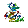
| ||||||||||||
|---|---|---|---|---|---|---|---|---|---|---|---|---|---|
| 1 |
| ||||||||||||
| Unit cell |
|
-Components
| #1: Protein | Mass: 39741.469 Da / Num. of mol.: 1 Source method: isolated from a genetically manipulated source Source: (gene. exp.) Homo sapiens (human) / Gene: CLK1, CLK / Plasmid: PLIC-SGC1 / Production host:  Escherichia coli (E. coli) / References: UniProt: P49759, dual-specificity kinase Escherichia coli (E. coli) / References: UniProt: P49759, dual-specificity kinase |
|---|---|
| #2: Chemical | ChemComp-V25 / 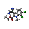  Mass: 338.189 Da / Num. of mol.: 1 / Source method: obtained synthetically / Formula: C15H13Cl2N3O2 Mass: 338.189 Da / Num. of mol.: 1 / Source method: obtained synthetically / Formula: C15H13Cl2N3O2 |
| #3: Water | ChemComp-HOH / Water Mass: 18.015 Da / Num. of mol.: 216 / Source method: isolated from a natural source / Formula: H2O Mass: 18.015 Da / Num. of mol.: 216 / Source method: isolated from a natural source / Formula: H2O |
| Has ligand of interest | N |
-Experimental details
-Experiment
| Experiment | Method: X-RAY DIFFRACTION / Number of used crystals: 1 |
|---|
- Sample preparation
| Crystal | Density Matthews: 2.55 Å3/Da / Density % sol: 51.78 % |
|---|---|
| Crystal grow | Temperature: 277 K / Method: vapor diffusion, hanging drop / pH: 7 / Details: 2.1 M SODIUM MALATE |
-Data collection
| Diffraction | Mean temperature: 100 K / Serial crystal experiment: N |
|---|---|
| Diffraction source | Source: SYNCHROTRON / Site: SLS  / Beamline: X10SA / Wavelength: 1.006029 Å / Beamline: X10SA / Wavelength: 1.006029 Å |
| Detector | Type: MARRESEARCH / Detector: CCD / Date: Jul 15, 2007 |
| Radiation | Protocol: SINGLE WAVELENGTH / Monochromatic (M) / Laue (L): M / Scattering type: x-ray |
| Radiation wavelength | Wavelength: 1.006029 Å / Relative weight: 1 |
| Reflection | Resolution: 1.8→30 Å / Num. obs: 36979 / % possible obs: 99.9 % / Redundancy: 3.8 % / Biso Wilson estimate: 25.61 Å2 / Rmerge(I) obs: 0.09 / Net I/σ(I): 11.6 |
| Reflection shell | Resolution: 1.8→1.92 Å / Redundancy: 3 % / Rmerge(I) obs: 0.613 / Mean I/σ(I) obs: 1.8 / Num. unique obs: 5380 / % possible all: 99.9 |
- Processing
| Software |
| ||||||||||||||||||||||||||||||||||||||||||||||||||||||||||||||||||||||||||||||||||||||||||||||||||
|---|---|---|---|---|---|---|---|---|---|---|---|---|---|---|---|---|---|---|---|---|---|---|---|---|---|---|---|---|---|---|---|---|---|---|---|---|---|---|---|---|---|---|---|---|---|---|---|---|---|---|---|---|---|---|---|---|---|---|---|---|---|---|---|---|---|---|---|---|---|---|---|---|---|---|---|---|---|---|---|---|---|---|---|---|---|---|---|---|---|---|---|---|---|---|---|---|---|---|---|
| Refinement | Method to determine structure: FOURIER SYNTHESIS Starting model: 2VAG Resolution: 1.8→29.27 Å / SU ML: 0.2533 / Cross valid method: FREE R-VALUE / σ(F): 1.38 / Phase error: 23.2524
| ||||||||||||||||||||||||||||||||||||||||||||||||||||||||||||||||||||||||||||||||||||||||||||||||||
| Solvent computation | Shrinkage radii: 0.9 Å / VDW probe radii: 1.11 Å | ||||||||||||||||||||||||||||||||||||||||||||||||||||||||||||||||||||||||||||||||||||||||||||||||||
| Displacement parameters | Biso mean: 35.75 Å2 | ||||||||||||||||||||||||||||||||||||||||||||||||||||||||||||||||||||||||||||||||||||||||||||||||||
| Refinement step | Cycle: LAST / Resolution: 1.8→29.27 Å
| ||||||||||||||||||||||||||||||||||||||||||||||||||||||||||||||||||||||||||||||||||||||||||||||||||
| Refine LS restraints |
| ||||||||||||||||||||||||||||||||||||||||||||||||||||||||||||||||||||||||||||||||||||||||||||||||||
| LS refinement shell |
| ||||||||||||||||||||||||||||||||||||||||||||||||||||||||||||||||||||||||||||||||||||||||||||||||||
| Refinement TLS params. | Method: refined / Origin x: 14.4105004445 Å / Origin y: 6.64873866416 Å / Origin z: 14.7635358718 Å
| ||||||||||||||||||||||||||||||||||||||||||||||||||||||||||||||||||||||||||||||||||||||||||||||||||
| Refinement TLS group | Selection details: (chain 'E' and resid 0 through 484) |
