 Movie
Movie Controller


+ Open data

- Basic information
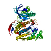
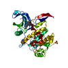
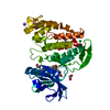
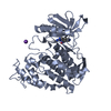
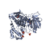
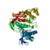
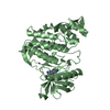
| Entry | Database: PDB / ID: 6yti | ||||||
|---|---|---|---|---|---|---|---|
| Title | CLK1 bound with ETH1610 (Cpd 17) | ||||||
 Components Components | Dual specificity protein kinase CLK1 | ||||||
 Keywords Keywords |  TRANSFERASE / Inhibitor / Complex / CLK1 / Structural Genomics / Structural Genomics Consortium / SGC TRANSFERASE / Inhibitor / Complex / CLK1 / Structural Genomics / Structural Genomics Consortium / SGC | ||||||
| Function / homology |  Function and homology informationdual-specificity kinase / regulation of RNA splicing / protein serine/threonine/tyrosine kinase activity / non-membrane spanning protein tyrosine kinase activity / protein tyrosine kinase activity / phosphorylation / protein serine kinase activity / protein serine/threonine kinase activity / ATP binding / nucleus Function and homology informationdual-specificity kinase / regulation of RNA splicing / protein serine/threonine/tyrosine kinase activity / non-membrane spanning protein tyrosine kinase activity / protein tyrosine kinase activity / phosphorylation / protein serine kinase activity / protein serine/threonine kinase activity / ATP binding / nucleusSimilarity search - Function | ||||||
| Biological species |  Homo sapiens (human) Homo sapiens (human) | ||||||
| Method | X-RAY DIFFRACTION / SYNCHROTRON / MOLECULAR REPLACEMENT / Resolution: 2.4 Å | ||||||
 Authors Authors | Schroeder, M. / Chaikuad, A. / Knapp, S. / Structural Genomics Consortium (SGC) | ||||||
 Citation Citation | Journal: J.Med.Chem. / Year: 2020 Title: DFG-1 Residue Controls Inhibitor Binding Mode and Affinity, Providing a Basis for Rational Design of Kinase Inhibitor Selectivity. Authors: Schroder, M. / Bullock, A.N. / Fedorov, O. / Bracher, F. / Chaikuad, A. / Knapp, S. | ||||||
| History |
|
- Structure visualization
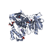
| Structure viewer | Molecule: MolmilJmol/JSmol |
|---|
- Downloads & links
-Download
| PDBx/mmCIF format | 6yti.cif.gz | 152.4 KB | Display | PDBx/mmCIF format |
|---|---|---|---|---|
| PDB format | pdb6yti.ent.gz | 118.6 KB | Display | PDB format |
| PDBx/mmJSON format | 6yti.json.gz | Tree view | PDBx/mmJSON format | |
| Others |  Other downloads Other downloads |
-Validation report
| Arichive directory | https://data.pdbj.org/pub/pdb/validation_reports/yt/6ytiftp://data.pdbj.org/pub/pdb/validation_reports/yt/6yti | HTTPS FTP |
|---|
-Related structure data
| Related structure data | 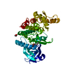 6ytaC 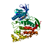 6ytdC 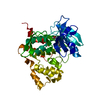 6yteC 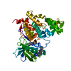 6ytgC  6ytwC  6ytyC 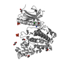 6yu1C  6z2vC 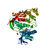 6zlnC 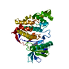 1z57S S: Starting model for refinement C: citing same article ( |
|---|---|
| Similar structure data |


-Links
 PDBj
PDBj- Assembly
| Deposited unit | 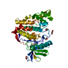
| ||||||||
|---|---|---|---|---|---|---|---|---|---|
| 1 |
| ||||||||
| Unit cell |
| ||||||||
| Components on special symmetry positions |
|
-Components
| #1: Protein | Mass: 39581.512 Da / Num. of mol.: 1 Source method: isolated from a genetically manipulated source Source: (gene. exp.) Homo sapiens (human) / Gene: CLK1, CLK / Production host:  Escherichia coli (E. coli) / References: UniProt: P49759, dual-specificity kinase Escherichia coli (E. coli) / References: UniProt: P49759, dual-specificity kinase | ||||||
|---|---|---|---|---|---|---|---|
| #2: Chemical | ChemComp-EDO / Ethylene glycol  Mass: 62.068 Da / Num. of mol.: 4 / Source method: obtained synthetically / Formula: C2H6O2 Mass: 62.068 Da / Num. of mol.: 4 / Source method: obtained synthetically / Formula: C2H6O2#3: Chemical | ChemComp-7A7 / | 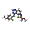  Mass: 383.399 Da / Num. of mol.: 1 / Source method: obtained synthetically / Formula: C18H14FN5O2S / Feature type: SUBJECT OF INVESTIGATION Mass: 383.399 Da / Num. of mol.: 1 / Source method: obtained synthetically / Formula: C18H14FN5O2S / Feature type: SUBJECT OF INVESTIGATION#4: Water | ChemComp-HOH / | Water Mass: 18.015 Da / Num. of mol.: 87 / Source method: isolated from a natural source / Formula: H2O Mass: 18.015 Da / Num. of mol.: 87 / Source method: isolated from a natural source / Formula: H2OHas ligand of interest | Y | |
-Experimental details
-Experiment
| Experiment | Method: X-RAY DIFFRACTION / Number of used crystals: 1 |
|---|
- Sample preparation
| Crystal | Density Matthews: 2.57 Å3/Da / Density % sol: 52.11 % |
|---|---|
| Crystal grow | Temperature: 277 K / Method: vapor diffusion, sitting drop / pH: 9.3 / Details: 29% PEG 6k, 0.1M bicine |
-Data collection
| Diffraction | Mean temperature: 100 K / Serial crystal experiment: N | |||||||||||||||||||||||||||
|---|---|---|---|---|---|---|---|---|---|---|---|---|---|---|---|---|---|---|---|---|---|---|---|---|---|---|---|---|
| Diffraction source | Source: SYNCHROTRON / Site: SLS  / Beamline: X06DA / Wavelength: 1 Å / Beamline: X06DA / Wavelength: 1 Å | |||||||||||||||||||||||||||
| Detector | Type: DECTRIS PILATUS 2M-F / Detector: PIXEL / Date: Mar 23, 2019 | |||||||||||||||||||||||||||
| Radiation | Protocol: SINGLE WAVELENGTH / Monochromatic (M) / Laue (L): M / Scattering type: x-ray | |||||||||||||||||||||||||||
| Radiation wavelength | Wavelength: 1 Å / Relative weight: 1 | |||||||||||||||||||||||||||
| Reflection | Resolution: 2.4→48.03 Å / Num. obs: 15727 / % possible obs: 99.4 % / Redundancy: 7 % / CC1/2: 0.996 / Rmerge(I) obs: 0.153 / Rpim(I) all: 0.062 / Rrim(I) all: 0.166 / Net I/σ(I): 10.1 | |||||||||||||||||||||||||||
| Reflection shell | Diffraction-ID: 1 / Redundancy: 6.9 %
|
- Processing
| Software |
| |||||||||||||||||||||||||||||||||||||||||||||||||||||||||||||||||||||||||||
|---|---|---|---|---|---|---|---|---|---|---|---|---|---|---|---|---|---|---|---|---|---|---|---|---|---|---|---|---|---|---|---|---|---|---|---|---|---|---|---|---|---|---|---|---|---|---|---|---|---|---|---|---|---|---|---|---|---|---|---|---|---|---|---|---|---|---|---|---|---|---|---|---|---|---|---|---|
| Refinement | Method to determine structure: MOLECULAR REPLACEMENT Starting model: 1z57 Resolution: 2.4→48.03 Å / Cor.coef. Fo:Fc: 0.946 / Cor.coef. Fo:Fc free: 0.912 / SU B: 20.464 / SU ML: 0.229 / SU R Cruickshank DPI: 0.412 / Cross valid method: THROUGHOUT / σ(F): 0 / ESU R: 0.412 / ESU R Free: 0.271 Details: HYDROGENS HAVE BEEN ADDED IN THE RIDING POSITIONS U VALUES : WITH TLS ADDED
| |||||||||||||||||||||||||||||||||||||||||||||||||||||||||||||||||||||||||||
| Solvent computation | Ion probe radii: 0.8 Å / Shrinkage radii: 0.8 Å / VDW probe radii: 1.2 Å | |||||||||||||||||||||||||||||||||||||||||||||||||||||||||||||||||||||||||||
| Displacement parameters | Biso max: 118.11 Å2 / Biso mean: 44.464 Å2 / Biso min: 26.41 Å2
| |||||||||||||||||||||||||||||||||||||||||||||||||||||||||||||||||||||||||||
| Refinement step | Cycle: final / Resolution: 2.4→48.03 Å
| |||||||||||||||||||||||||||||||||||||||||||||||||||||||||||||||||||||||||||
| Refine LS restraints |
| |||||||||||||||||||||||||||||||||||||||||||||||||||||||||||||||||||||||||||
| LS refinement shell | Resolution: 2.4→2.462 Å / Rfactor Rfree error: 0 / Total num. of bins used: 20
| |||||||||||||||||||||||||||||||||||||||||||||||||||||||||||||||||||||||||||
| Refinement TLS params. | Method: refined / Refine-ID: X-RAY DIFFRACTION
| |||||||||||||||||||||||||||||||||||||||||||||||||||||||||||||||||||||||||||
| Refinement TLS group |
|
