 Movie
Movie Controller

[English] 日本語
 Yorodumi
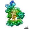
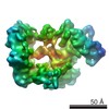
- PDB-6ss4: Structure of arginase-2 in complex with the inhibitory human anti... -
+ Open data

- Basic information
| Entry | Database: PDB / ID: 6ss4 | ||||||
|---|---|---|---|---|---|---|---|
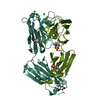
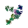
| Title | Structure of arginase-2 in complex with the inhibitory human antigen-binding fragment Fab C0021181 | ||||||
 Components Components |
| ||||||
 Keywords Keywords |  PROTEIN BINDING / arginase-2 inhibitor / IgG / antigen-binding fragment PROTEIN BINDING / arginase-2 inhibitor / IgG / antigen-binding fragment | ||||||
| Function / homology |  Function and homology information Function and homology informationnegative regulation of chemokine (C-C motif) ligand 4 production / negative regulation of activated CD8-positive, alpha-beta T cell apoptotic process / negative regulation of defense response to bacterium / negative regulation of macrophage inflammatory protein 1 alpha production / negative regulation of type 2 immune response / negative regulation of interleukin-13 production / negative regulation of chemokine (C-C motif) ligand 5 production / regulation of interleukin-1 beta production / Urea cycle / arginine catabolic process to ornithine ...negative regulation of chemokine (C-C motif) ligand 4 production / negative regulation of activated CD8-positive, alpha-beta T cell apoptotic process / negative regulation of defense response to bacterium / negative regulation of macrophage inflammatory protein 1 alpha production / negative regulation of type 2 immune response / negative regulation of interleukin-13 production / negative regulation of chemokine (C-C motif) ligand 5 production / regulation of interleukin-1 beta production / Urea cycle / arginine catabolic process to ornithine / arginase / arginase activity / urea cycle / negative regulation of CD4-positive, alpha-beta T cell proliferation / negative regulation of interleukin-17 production / regulation of reactive oxygen species biosynthetic process / ureteric bud development / negative regulation of tumor necrosis factor production / striated muscle contraction / nitric oxide biosynthetic process / positive regulation of cellular senescence / manganese ion binding / adaptive immune response / mitochondrial matrix / innate immune response / mitochondrion / cytoplasmSimilarity search - Function | ||||||
| Biological species |  Homo sapiens (human) Homo sapiens (human) | ||||||
| Method | X-RAY DIFFRACTION / SYNCHROTRON / MOLECULAR REPLACEMENT / Resolution: 2.9 Å | ||||||
 Authors Authors | Burschowsky, D. / Addyman, A. / Fiedler, S. / Groves, M. / Haynes, S. / Seewooruthun, C. / Carr, M. | ||||||
| Funding support |  United Kingdom, 1items United Kingdom, 1items
| ||||||
 Citation Citation | Journal: Mabs Title: Structural and functional characterization of C0021158, a high-affinity monoclonal antibody that inhibits Arginase 2 function via a novel non-competitive mechanism of action. Authors: Austin, M. / Burschowsky, D. / Chan, D.T.Y. / Jenkinson, L. / Haynes, S. / Diamandakis, A. / Seewooruthun, C. / Addyman, A. / Fiedler, S. / Ryman, S. / Whitehouse, J. / Slater, L.H. / ...Authors: Austin, M. / Burschowsky, D. / Chan, D.T.Y. / Jenkinson, L. / Haynes, S. / Diamandakis, A. / Seewooruthun, C. / Addyman, A. / Fiedler, S. / Ryman, S. / Whitehouse, J. / Slater, L.H. / Hadjinicolaou, A.V. / Gileadi, U. / Gowans, E. / Shibata, Y. / Barnard, M. / Kaserer, T. / Sharma, P. / Luheshi, N.M. / Wilkinson, R.W. / Vaughan, T.J. / Holt, S.V. / Cerundolo, V. / Carr, M.D. / Groves, M.A.T. | ||||||
| History |
|
- Structure visualization
| Structure viewer | Molecule: MolmilJmol/JSmol |
|---|
- Downloads & links
-Download
| PDBx/mmCIF format | 6ss4.cif.gz | 504.6 KB | Display | PDBx/mmCIF format |
|---|---|---|---|---|
| PDB format | pdb6ss4.ent.gz | Display | PDB format | |
| PDBx/mmJSON format | 6ss4.json.gz | Tree view | PDBx/mmJSON format | |
| Others |  Other downloads Other downloads |
-Validation report
| Arichive directory | https://data.pdbj.org/pub/pdb/validation_reports/ss/6ss4ftp://data.pdbj.org/pub/pdb/validation_reports/ss/6ss4 | HTTPS FTP |
|---|
-Related structure data
| Related structure data | 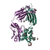 6srvC 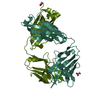 6srxC 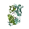 6ss0SC 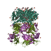 6ss2C  6tulC 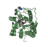 4hzeS C: citing same article ( S: Starting model for refinement |
|---|---|
| Similar structure data |


-Links
 PDBj
PDBj


- Assembly
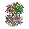
| Deposited unit | 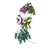
| ||||||||
|---|---|---|---|---|---|---|---|---|---|
| 1 | 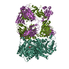
| ||||||||
| Unit cell |
|
-Components
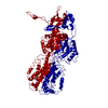
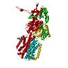
| #1: Protein | / Arginase II / Kidney-type arginase / Non-hepatic arginase / Type II arginase Mass: 37084.859 Da / Num. of mol.: 1 Source method: isolated from a genetically manipulated source Source: (gene. exp.) Homo sapiens (human) / Gene: ARG2 / Production host:  Escherichia coli BL21(DE3) (bacteria) / References: UniProt: P78540, arginase Escherichia coli BL21(DE3) (bacteria) / References: UniProt: P78540, arginase | ||||
|---|---|---|---|---|---|
| #2: Antibody | Mass: 24819.836 Da / Num. of mol.: 1 Source method: isolated from a genetically manipulated source Details: Amino acids are numbered according to the Kabat numbering scheme. Source: (gene. exp.) Homo sapiens (human) / Plasmid: pEU1.3 fab / Cell line (production host): CHO / Production host:   Cricetulus griseus (Chinese hamster) / Variant (production host): ExpiCHO Cricetulus griseus (Chinese hamster) / Variant (production host): ExpiCHO | ||||
| #3: Antibody | Mass: 23124.482 Da / Num. of mol.: 1 Source method: isolated from a genetically manipulated source Details: Amino acids are numbered according to the Kabat numbering scheme. Residue 10 would not be present in the Kabat scheme, but due to problems with non-continuous numbering in L-peptides, we ...Details: Amino acids are numbered according to the Kabat numbering scheme. Residue 10 would not be present in the Kabat scheme, but due to problems with non-continuous numbering in L-peptides, we included it. Therefore, residues 2-10 should be read as 1-9, per Kabat. Source: (gene. exp.) Homo sapiens (human) / Plasmid: pEU1.3 fab / Cell line (production host): CHO / Production host: Cricetulus griseus (Chinese hamster) / Variant (production host): ExpiCHO | ||||
| #4: Chemical |   Mass: 54.938 Da / Num. of mol.: 2 / Source method: obtained synthetically / Formula: Mn Mass: 54.938 Da / Num. of mol.: 2 / Source method: obtained synthetically / Formula: Mn#5: Chemical | ChemComp-PO4 / | Phosphate  Mass: 94.971 Da / Num. of mol.: 1 / Source method: obtained synthetically / Formula: PO4 Mass: 94.971 Da / Num. of mol.: 1 / Source method: obtained synthetically / Formula: PO4Has ligand of interest | N | |
-Experimental details
-Experiment
| Experiment | Method: X-RAY DIFFRACTION / Number of used crystals: 1 |
|---|
- Sample preparation
| Crystal | Density Matthews: 4.25 Å3/Da / Density % sol: 71.07 % |
|---|---|
| Crystal grow | Temperature: 291 K / Method: vapor diffusion, sitting drop / pH: 5 Details: 100 mM MMT pH 5.0 (malic acid, MES, tris) 20% glycerol 10% PEG4000 15 mM NaNO3 15 mM Na2HPO4 15 mM (NH4)2SO4 |
-Data collection
| Diffraction | Mean temperature: 100 K / Serial crystal experiment: N | ||||||||||||||||||
|---|---|---|---|---|---|---|---|---|---|---|---|---|---|---|---|---|---|---|---|
| Diffraction source | Source: SYNCHROTRON / Site: BESSY  / Beamline: 14.1 / Wavelength: 0.9184 Å / Beamline: 14.1 / Wavelength: 0.9184 Å | ||||||||||||||||||
| Detector | Type: DECTRIS PILATUS 6M-F / Detector: PIXEL / Date: May 12, 2018 | ||||||||||||||||||
| Radiation | Protocol: SINGLE WAVELENGTH / Monochromatic (M) / Laue (L): M / Scattering type: x-ray | ||||||||||||||||||
| Radiation wavelength | Wavelength: 0.9184 Å / Relative weight: 1 | ||||||||||||||||||
| Reflection twin |
| ||||||||||||||||||
| Reflection | Resolution: 2.9→44.98 Å / Num. obs: 32384 / % possible obs: 100 % / Redundancy: 20.2 % / CC1/2: 1 / Rmerge(I) obs: 0.35 / Net I/σ(I): 11 | ||||||||||||||||||
| Reflection shell | Resolution: 2.9→3.06 Å / Redundancy: 20.8 % / Rmerge(I) obs: 5.19 / Mean I/σ(I) obs: 0.7 / Num. unique obs: 4672 / CC1/2: 0.32 / % possible all: 100 |
- Processing
| Software |
| |||||||||||||||||||||||||||||||||||||||||||||||||||||||||||||||||||||||||||||||||||||||||||||||||||||||||||||||||||||||||||||||||||||||||||||||||||||||||||
|---|---|---|---|---|---|---|---|---|---|---|---|---|---|---|---|---|---|---|---|---|---|---|---|---|---|---|---|---|---|---|---|---|---|---|---|---|---|---|---|---|---|---|---|---|---|---|---|---|---|---|---|---|---|---|---|---|---|---|---|---|---|---|---|---|---|---|---|---|---|---|---|---|---|---|---|---|---|---|---|---|---|---|---|---|---|---|---|---|---|---|---|---|---|---|---|---|---|---|---|---|---|---|---|---|---|---|---|---|---|---|---|---|---|---|---|---|---|---|---|---|---|---|---|---|---|---|---|---|---|---|---|---|---|---|---|---|---|---|---|---|---|---|---|---|---|---|---|---|---|---|---|---|---|---|---|---|
| Refinement | Method to determine structure: MOLECULAR REPLACEMENT Starting model: 4HZE,6SS0 Resolution: 2.9→44.98 Å / Cor.coef. Fo:Fc: 0.923 / Cor.coef. Fo:Fc free: 0.892 / WRfactor Rfree: 0.219 / WRfactor Rwork: 0.176 / SU B: 25.779 / SU ML: 0.224 / Average fsc free: 0.9648 / Average fsc work: 0.981 / Cross valid method: FREE R-VALUE / ESU R: 0.137 / ESU R Free: 0.085 Details: Hydrogens have been added in their riding positions
| |||||||||||||||||||||||||||||||||||||||||||||||||||||||||||||||||||||||||||||||||||||||||||||||||||||||||||||||||||||||||||||||||||||||||||||||||||||||||||
| Solvent computation | Ion probe radii: 0.8 Å / Shrinkage radii: 0.8 Å / VDW probe radii: 1.2 Å / Solvent model: MASK BULK SOLVENT | |||||||||||||||||||||||||||||||||||||||||||||||||||||||||||||||||||||||||||||||||||||||||||||||||||||||||||||||||||||||||||||||||||||||||||||||||||||||||||
| Displacement parameters | Biso mean: 79.623 Å2
| |||||||||||||||||||||||||||||||||||||||||||||||||||||||||||||||||||||||||||||||||||||||||||||||||||||||||||||||||||||||||||||||||||||||||||||||||||||||||||
| Refinement step | Cycle: LAST / Resolution: 2.9→44.98 Å
| |||||||||||||||||||||||||||||||||||||||||||||||||||||||||||||||||||||||||||||||||||||||||||||||||||||||||||||||||||||||||||||||||||||||||||||||||||||||||||
| Refine LS restraints |
| |||||||||||||||||||||||||||||||||||||||||||||||||||||||||||||||||||||||||||||||||||||||||||||||||||||||||||||||||||||||||||||||||||||||||||||||||||||||||||
| LS refinement shell |
| |||||||||||||||||||||||||||||||||||||||||||||||||||||||||||||||||||||||||||||||||||||||||||||||||||||||||||||||||||||||||||||||||||||||||||||||||||||||||||
| Refinement TLS params. | Method: refined / Refine-ID: X-RAY DIFFRACTION
| |||||||||||||||||||||||||||||||||||||||||||||||||||||||||||||||||||||||||||||||||||||||||||||||||||||||||||||||||||||||||||||||||||||||||||||||||||||||||||
| Refinement TLS group |
|
