- EMDB-4142: Cryo-EM structure of the E. coli replicative DNA polymerase-clamp... -
+
Open data
ID or keywords:
Loading...
-
Basic information
Entry
Database: EMDB / ID: EMD-4142
Title
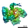
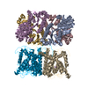
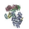
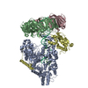
Cryo-EM structure of the E. coli replicative DNA polymerase-clamp-exonuclase-theta complex bound to DNA in the editing mode
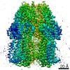
Map data
Related to EMD-4142 Local alignment after signal subtraction of the beta subunit
Sample
Complex: DNA polyerase III alpha, beta, epsilon, theta complex with mismatched DNA duplex
Complex: DNA polyerase III alpha, epsilon, theta complex with mismatched DNA duplex
Function / homology
Function and homology information
DNA polymerase III, core complex / Hda-beta clamp complex / bacterial-type DNA replication / replication inhibiting complex / DNA replication proofreading / DNA polymerase III complex / lagging strand elongation / replisome / regulation of DNA-templated DNA replication initiation / DNA strand elongation involved in DNA replication ...DNA polymerase III, core complex / Hda-beta clamp complex / bacterial-type DNA replication / replication inhibiting complex / DNA replication proofreading / DNA polymerase III complex / lagging strand elongation / replisome / regulation of DNA-templated DNA replication initiation / DNA strand elongation involved in DNA replication / exonuclease activity / leading strand elongation / error-prone translesion synthesis / negative regulation of DNA-templated DNA replication initiation / 3'-5' exonuclease activity / DNA-templated DNA replication / DNA-directed DNA polymerase / DNA-directed DNA polymerase activity / DNA damage response / protein homodimerization activity / DNA binding / identical protein binding / metal ion binding / cytosol / cytoplasm Similarity search - Function
DNA polymerase III-theta, bacterial / DNA polymerase III-theta superfamily / DNA polymerase III, theta subunit / : / : / DNA polymerase III subunit alpha, C-terminal domain / DNA polymerase 3, epsilon subunit / DNA polymerase III epsilon subunit, exonuclease domain / Bacterial DNA polymerase III alpha subunit, thumb domain / DNA polymerase III, alpha subunit ...DNA polymerase III-theta, bacterial / DNA polymerase III-theta superfamily / DNA polymerase III, theta subunit / : / : / DNA polymerase III subunit alpha, C-terminal domain / DNA polymerase 3, epsilon subunit / DNA polymerase III epsilon subunit, exonuclease domain / Bacterial DNA polymerase III alpha subunit, thumb domain / DNA polymerase III, alpha subunit / Bacterial DNA polymerase III, alpha subunit, NTPase domain / DNA polymerase, helix-hairpin-helix motif / DNA polymerase III alpha subunit finger domain / Bacterial DNA polymerase III alpha NTPase domain / Helix-hairpin-helix motif / Bacterial DNA polymerase III alpha subunit finger domain / PHP domain / PHP domain / Polymerase/histidinol phosphatase, N-terminal / DNA polymerase alpha chain like domain / Polymerase/histidinol phosphatase-like / Exonuclease / DNA polymerase III, beta sliding clamp / DNA polymerase III, beta sliding clamp, N-terminal / DNA polymerase III, beta sliding clamp, C-terminal / DNA polymerase III, beta sliding clamp, central / DNA polymerase III beta subunit, N-terminal domain / DNA polymerase III beta subunit, central domain / DNA polymerase III beta subunit, C-terminal domain / DNA polymerase III beta subunit / Exonuclease, RNase T/DNA polymerase III / EXOIII / OB-fold nucleic acid binding domain, AA-tRNA synthetase-type / OB-fold nucleic acid binding domain / : / Ribonuclease H superfamily / Ribonuclease H-like superfamily / Nucleic acid-binding, OB-fold Similarity search - Domain/homology
DNA polymerase III subunit epsilon / Beta sliding clamp / DNA polymerase III subunit theta / DNA polymerase III subunit alpha Similarity search - Component
Biological species
Escherichia coli (E. coli)
Method
single particle reconstruction / cryo EM / Resolution: 6.7 Å
Journal: Nat Struct Mol Biol / Year: 2017 Title: Self-correcting mismatches during high-fidelity DNA replication. Authors: Rafael Fernandez-Leiro / Julian Conrad / Ji-Chun Yang / Stefan M V Freund / Sjors H W Scheres / Meindert H Lamers / Abstract: Faithful DNA replication is essential to all forms of life and depends on the action of 3'-5' exonucleases that remove misincorporated nucleotides from the newly synthesized strand. However, how the ...Faithful DNA replication is essential to all forms of life and depends on the action of 3'-5' exonucleases that remove misincorporated nucleotides from the newly synthesized strand. However, how the DNA is transferred from the polymerase to the exonuclease active site is not known. Here we present the cryo-EM structure of the editing mode of the catalytic core of the Escherichia coli replisome, revealing a dramatic distortion of the DNA whereby the polymerase thumb domain acts as a wedge that separates the two DNA strands. Importantly, NMR analysis of the DNA substrate shows that the presence of a mismatch increases the fraying of the DNA, thus enabling it to reach the exonuclease active site. Therefore the mismatch corrects itself, whereas the exonuclease subunit plays a passive role. Hence, our work provides unique insights into high-fidelity replication and establishes a new paradigm for the correction of misincorporated nucleotides.
History
Deposition
Oct 12, 2016
-
Header (metadata) release
Oct 26, 2016
-
Map release
Jan 18, 2017
-
Update
Aug 30, 2017
-
Current status
Aug 30, 2017
Processing site: PDBe / Status: Released
-
Structure visualization
Movie
Surface view with section colored by density value
Supramolecule #2: DNA polyerase III alpha, epsilon, theta complex with mismatched D...
Supramolecule
Name: DNA polyerase III alpha, epsilon, theta complex with mismatched DNA duplex type: complex / ID: 2 / Parent: 1 / Macromolecule list: #1, #3-#6 Details: Map obtained after signal subtraction of the beta subunit and alignment of the remaining parts. Final reconstruction obtained with non-subtracted images and angles from local alignment
Source (natural)
Organism: Escherichia coli (E. coli) / Strain: K12
Cryogen name: ETHANE / Chamber humidity: 100 % / Chamber temperature: 277 K / Instrument: FEI VITROBOT MARK IV Details: Prior to sample preparation 0.1 volumes of 0.05% Tween 20 were added to the sample 3 microliters were pipetted onto the grid and blotted for 4 seconds.
Details
Sample was run over a gel filtration column prior to vitrification
-
Electron microscopy
Microscope
FEI TITAN KRIOS
Electron beam
Acceleration voltage: 300 kV / Electron source: FIELD EMISSION GUN
Algorithm: FOURIER SPACE / Resolution.type: BY AUTHOR / Resolution: 6.7 Å / Resolution method: FSC 0.143 CUT-OFF / Software - Name: RELION (ver. 2) / Number images used: 15616
FSC plot (resolution estimation)
-
Atomic model buiding 1
Details
The cryo-EM structure of the PolIIIalpha-clamp-exonuclease complex in the polymerase mode (PDB code: 5FKW)1 was used as a starting model, and the NMR structure of theta bound to the ? catalytic domain (PDB code: 2XY8)13 was used to place ? into the cryo-EM map. The model was manually adjusted in Coot35 and geometry of the protein optimized in Refmac536 using DNA-specific restraints generated in LibG36
Refinement
Protocol: OTHER
+
About Yorodumi
-
News
-
Feb 9, 2022. New format data for meta-information of EMDB entries
New format data for meta-information of EMDB entries
Version 3 of the EMDB header file is now the official format.
The previous official version 1.9 will be removed from the archive.
In the structure databanks used in Yorodumi, some data are registered as the other names, "COVID-19 virus" and "2019-nCoV". Here are the details of the virus and the list of structure data.
Jan 31, 2019. EMDB accession codes are about to change! (news from PDBe EMDB page)
EMDB accession codes are about to change! (news from PDBe EMDB page)
The allocation of 4 digits for EMDB accession codes will soon come to an end. Whilst these codes will remain in use, new EMDB accession codes will include an additional digit and will expand incrementally as the available range of codes is exhausted. The current 4-digit format prefixed with “EMD-” (i.e. EMD-XXXX) will advance to a 5-digit format (i.e. EMD-XXXXX), and so on. It is currently estimated that the 4-digit codes will be depleted around Spring 2019, at which point the 5-digit format will come into force.
The EM Navigator/Yorodumi systems omit the EMD- prefix.
Related info.:Q: What is EMD? / ID/Accession-code notation in Yorodumi/EM Navigator
Yorodumi is a browser for structure data from EMDB, PDB, SASBDB, etc.
This page is also the successor to EM Navigator detail page, and also detail information page/front-end page for Omokage search.
The word "yorodu" (or yorozu) is an old Japanese word meaning "ten thousand". "mi" (miru) is to see.
Related info.:EMDB / PDB / SASBDB / Comparison of 3 databanks / Yorodumi Search / Aug 31, 2016. New EM Navigator & Yorodumi / Yorodumi Papers / Jmol/JSmol / Function and homology information / Changes in new EM Navigator and Yorodumi
 Movie
Movie Controller

 Yorodumi
 Open data
 Basic information
 Map data
 Sample
 Function and homology information
 DNA polymerase III, core complex / Hda-beta clamp complex / bacterial-type DNA replication / replication inhibiting complex / DNA replication proofreading /
 Authors
 Citation
 Structure visualization
 Downloads & links
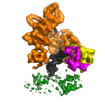 emd_4142.png
 http://ftp.pdbj.org/pub/emdb/structures/EMD-4142


 Z
Z Y
Y X
X


 Sample components
 Processing
 Electron microscopy