 Movie
Movie Controller

[English] 日本語
 Yorodumi
- PDB-6smp: AntDE:AntF (holo): type II PKS acyl-carrier protein in complex wi... -
+ Open data

- Basic information
| Entry | Database: PDB / ID: 6smp | ||||||
|---|---|---|---|---|---|---|---|
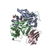
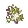
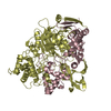
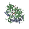
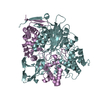
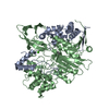
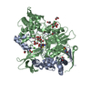
| Title | AntDE:AntF (holo): type II PKS acyl-carrier protein in complex with its ketosynthase bound to the hexaketide | ||||||
 Components Components |
| ||||||
 Keywords Keywords |  PROTEIN BINDING / Natural Product Biosynthesis / Polyketides / Minimal PKS System / Anthraquinone / Chain Elongation / Catalysis PROTEIN BINDING / Natural Product Biosynthesis / Polyketides / Minimal PKS System / Anthraquinone / Chain Elongation / Catalysis | ||||||
| Function / homology |  Function and homology information Function and homology informationacyltransferase activity, transferring groups other than amino-acyl groups Similarity search - Function | ||||||
| Biological species |  Photorhabdus luminescens (bacteria)Photorhabdus luminescens subsp. laumondii (bacteria) Photorhabdus luminescens (bacteria)Photorhabdus luminescens subsp. laumondii (bacteria) | ||||||
| Method | X-RAY DIFFRACTION / SYNCHROTRON / MOLECULAR REPLACEMENT / Resolution: 2.9 Å | ||||||
 Authors Authors | Braeuer, A. / Zhou, Q. / Grammbitter, G.L.C. / Schmalhofer, M. / Ruehl, M. / Kaila, V.R.I. / Bode, H. / Groll, M. | ||||||
 Citation Citation | Journal: Nat.Chem. / Year: 2020 Title: Structural snapshots of the minimal PKS system responsible for octaketide biosynthesis. Authors: Brauer, A. / Zhou, Q. / Grammbitter, G.L.C. / Schmalhofer, M. / Ruhl, M. / Kaila, V.R.I. / Bode, H.B. / Groll, M. | ||||||
| History |
|
- Structure visualization
| Structure viewer | Molecule: MolmilJmol/JSmol |
|---|
- Downloads & links
-Download
| PDBx/mmCIF format | 6smp.cif.gz | 1.2 MB | Display | PDBx/mmCIF format |
|---|---|---|---|---|
| PDB format | pdb6smp.ent.gz | 1 MB | Display | PDB format |
| PDBx/mmJSON format | 6smp.json.gz | Tree view | PDBx/mmJSON format | |
| Others |  Other downloads Other downloads |
-Validation report
| Arichive directory | https://data.pdbj.org/pub/pdb/validation_reports/sm/6smpftp://data.pdbj.org/pub/pdb/validation_reports/sm/6smp | HTTPS FTP |
|---|
-Related structure data
| Related structure data |  6sm4C  6sm6C  6smdC  6smoSC C: citing same article ( S: Starting model for refinement |
|---|---|
| Similar structure data |


-Links
 PDBj
PDBj

- Assembly
| Deposited unit | 
| ||||||||
|---|---|---|---|---|---|---|---|---|---|
| 1 | 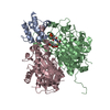
| ||||||||
| 2 | 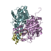
| ||||||||
| 3 | 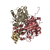
| ||||||||
| 4 | 
| ||||||||
| Unit cell |
|
-Components
| #1: Protein | Mass: 10850.877 Da / Num. of mol.: 4 Source method: isolated from a genetically manipulated source Source: (gene. exp.) Photorhabdus luminescens (bacteria) / Gene: C6H68_21855 / Production host: Escherichia coli BL21(DE3) (bacteria) / References: UniProt: A0A2S8QL96, UniProt: Q7MZT5*PLUS#2: Protein | Mass: 47350.023 Da / Num. of mol.: 4 Source method: isolated from a genetically manipulated source Source: (gene. exp.) Photorhabdus luminescens subsp. laumondii (strain DSM 15139 / CIP 105565 / TT01) (bacteria)Strain: DSM 15139 / CIP 105565 / TT01 / Gene: plu4191 / Production host: Escherichia coli BL21(DE3) (bacteria) / References: UniProt: Q7MZT3#3: Protein | Mass: 40787.746 Da / Num. of mol.: 4 Source method: isolated from a genetically manipulated source Source: (gene. exp.) Photorhabdus luminescens subsp. laumondii (strain DSM 15139 / CIP 105565 / TT01) (bacteria)Strain: DSM 15139 / CIP 105565 / TT01 / Gene: plu4190 / Production host: Escherichia coli BL21(DE3) (bacteria) / References: UniProt: Q7MZT4#4: Chemical | ChemComp-LLE / (   Mass: 614.600 Da / Num. of mol.: 4 / Source method: obtained synthetically / Formula: C23H39N2O13PS Mass: 614.600 Da / Num. of mol.: 4 / Source method: obtained synthetically / Formula: C23H39N2O13PS#5: Water | ChemComp-HOH / | Water Mass: 18.015 Da / Num. of mol.: 81 / Source method: isolated from a natural source / Formula: H2O Mass: 18.015 Da / Num. of mol.: 81 / Source method: isolated from a natural source / Formula: H2OHas ligand of interest | Y | |
|---|
-Experimental details
-Experiment
| Experiment | Method: X-RAY DIFFRACTION / Number of used crystals: 1 |
|---|
- Sample preparation
| Crystal | Density Matthews: 2.99 Å3/Da / Density % sol: 58.84 % |
|---|---|
| Crystal grow | Temperature: 293 K / Method: vapor diffusion, sitting drop / pH: 6 / Details: 0.1 M BIS-TRIS, 0.5 M (NH4)2SO4, 30% PEG 3350 |
-Data collection
| Diffraction | Mean temperature: 100 K / Serial crystal experiment: N |
|---|---|
| Diffraction source | Source: SYNCHROTRON / Site: SLS  / Beamline: X06SA / Wavelength: 1 Å / Beamline: X06SA / Wavelength: 1 Å |
| Detector | Type: DECTRIS EIGER X 16M / Detector: PIXEL / Date: Oct 15, 2016 |
| Radiation | Protocol: SINGLE WAVELENGTH / Monochromatic (M) / Laue (L): M / Scattering type: x-ray |
| Radiation wavelength | Wavelength: 1 Å / Relative weight: 1 |
| Reflection | Resolution: 2.9→50 Å / Num. obs: 92717 / % possible obs: 97.9 % / Redundancy: 3.1 % / CC1/2: 0.993 / Rmerge(I) obs: 0.097 / Net I/σ(I): 11.4 |
| Reflection shell | Resolution: 2.9→3 Å / Rmerge(I) obs: 0.543 / Mean I/σ(I) obs: 2.4 / Num. unique obs: 8962 / CC1/2: 0.734 / % possible all: 98.4 |
- Processing
| Software |
| |||||||||||||||||||||||||||||||||||||||||||||||||||||||||||||||||||||||||||||||||||||||||||||||||||||||||||||||||||||||||||||||||||||||||||||||||||||||||||||||||||||||||||||||||||||||||||||||||||||||||||||||||||||||||||||||||||||||||||||||||||||||||||||||||||||||||||||||||||||||||||||||||||||||||||||||||||||||||||||||||||||
|---|---|---|---|---|---|---|---|---|---|---|---|---|---|---|---|---|---|---|---|---|---|---|---|---|---|---|---|---|---|---|---|---|---|---|---|---|---|---|---|---|---|---|---|---|---|---|---|---|---|---|---|---|---|---|---|---|---|---|---|---|---|---|---|---|---|---|---|---|---|---|---|---|---|---|---|---|---|---|---|---|---|---|---|---|---|---|---|---|---|---|---|---|---|---|---|---|---|---|---|---|---|---|---|---|---|---|---|---|---|---|---|---|---|---|---|---|---|---|---|---|---|---|---|---|---|---|---|---|---|---|---|---|---|---|---|---|---|---|---|---|---|---|---|---|---|---|---|---|---|---|---|---|---|---|---|---|---|---|---|---|---|---|---|---|---|---|---|---|---|---|---|---|---|---|---|---|---|---|---|---|---|---|---|---|---|---|---|---|---|---|---|---|---|---|---|---|---|---|---|---|---|---|---|---|---|---|---|---|---|---|---|---|---|---|---|---|---|---|---|---|---|---|---|---|---|---|---|---|---|---|---|---|---|---|---|---|---|---|---|---|---|---|---|---|---|---|---|---|---|---|---|---|---|---|---|---|---|---|---|---|---|---|---|---|---|---|---|---|---|---|---|---|---|---|---|---|---|---|---|---|---|---|---|---|---|---|---|---|---|---|---|---|---|---|---|---|---|---|---|---|---|---|---|---|---|---|---|---|---|---|---|---|---|---|---|---|---|---|---|---|---|---|---|---|---|---|
| Refinement | Method to determine structure: MOLECULAR REPLACEMENT Starting model: 6SMO Resolution: 2.9→15 Å / Cor.coef. Fo:Fc: 0.919 / Cor.coef. Fo:Fc free: 0.896 / SU B: 44.249 / SU ML: 0.357 / Cross valid method: THROUGHOUT / σ(F): 0 / ESU R Free: 0.417 / Details: U VALUES : WITH TLS ADDED
| |||||||||||||||||||||||||||||||||||||||||||||||||||||||||||||||||||||||||||||||||||||||||||||||||||||||||||||||||||||||||||||||||||||||||||||||||||||||||||||||||||||||||||||||||||||||||||||||||||||||||||||||||||||||||||||||||||||||||||||||||||||||||||||||||||||||||||||||||||||||||||||||||||||||||||||||||||||||||||||||||||||
| Solvent computation | Ion probe radii: 0.8 Å / Shrinkage radii: 0.8 Å / VDW probe radii: 1.2 Å | |||||||||||||||||||||||||||||||||||||||||||||||||||||||||||||||||||||||||||||||||||||||||||||||||||||||||||||||||||||||||||||||||||||||||||||||||||||||||||||||||||||||||||||||||||||||||||||||||||||||||||||||||||||||||||||||||||||||||||||||||||||||||||||||||||||||||||||||||||||||||||||||||||||||||||||||||||||||||||||||||||||
| Displacement parameters | Biso max: 149.74 Å2 / Biso mean: 61.457 Å2 / Biso min: 24.64 Å2
| |||||||||||||||||||||||||||||||||||||||||||||||||||||||||||||||||||||||||||||||||||||||||||||||||||||||||||||||||||||||||||||||||||||||||||||||||||||||||||||||||||||||||||||||||||||||||||||||||||||||||||||||||||||||||||||||||||||||||||||||||||||||||||||||||||||||||||||||||||||||||||||||||||||||||||||||||||||||||||||||||||||
| Refinement step | Cycle: final / Resolution: 2.9→15 Å
| |||||||||||||||||||||||||||||||||||||||||||||||||||||||||||||||||||||||||||||||||||||||||||||||||||||||||||||||||||||||||||||||||||||||||||||||||||||||||||||||||||||||||||||||||||||||||||||||||||||||||||||||||||||||||||||||||||||||||||||||||||||||||||||||||||||||||||||||||||||||||||||||||||||||||||||||||||||||||||||||||||||
| Refine LS restraints |
| |||||||||||||||||||||||||||||||||||||||||||||||||||||||||||||||||||||||||||||||||||||||||||||||||||||||||||||||||||||||||||||||||||||||||||||||||||||||||||||||||||||||||||||||||||||||||||||||||||||||||||||||||||||||||||||||||||||||||||||||||||||||||||||||||||||||||||||||||||||||||||||||||||||||||||||||||||||||||||||||||||||
| LS refinement shell | Resolution: 2.9→2.972 Å / Rfactor Rfree error: 0 / Total num. of bins used: 20
| |||||||||||||||||||||||||||||||||||||||||||||||||||||||||||||||||||||||||||||||||||||||||||||||||||||||||||||||||||||||||||||||||||||||||||||||||||||||||||||||||||||||||||||||||||||||||||||||||||||||||||||||||||||||||||||||||||||||||||||||||||||||||||||||||||||||||||||||||||||||||||||||||||||||||||||||||||||||||||||||||||||
| Refinement TLS params. | Method: refined / Refine-ID: X-RAY DIFFRACTION
| |||||||||||||||||||||||||||||||||||||||||||||||||||||||||||||||||||||||||||||||||||||||||||||||||||||||||||||||||||||||||||||||||||||||||||||||||||||||||||||||||||||||||||||||||||||||||||||||||||||||||||||||||||||||||||||||||||||||||||||||||||||||||||||||||||||||||||||||||||||||||||||||||||||||||||||||||||||||||||||||||||||
| Refinement TLS group |
|
