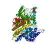
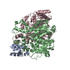
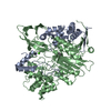
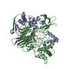
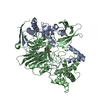
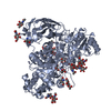
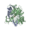
[English] 日本語
Yorodumi
- PDB-4pem: Crystal Structure of S1G mutant of Penicillin G Acylase from Kluy... -

+
Open data


ID or keywords:

Loading...

-
Basic information

Entry
Database: PDB / ID: 4pem
TitleCrystal Structure of S1G mutant of Penicillin G Acylase from Kluyvera citrophila
Components
  • Penicillin G acylase Alpha
  • Penicillin G acylase Beta
KeywordsHYDROLASE / Ntn Hydrolase / PGA / Slow processing Mutant
Function / homology
Function and homology information


hydrolase activity, acting on carbon-nitrogen (but not peptide) bonds, in linear amides / antibiotic biosynthetic process / metal ion binding
Similarity search - Function
Penicillin G acylase, C-terminal / Penicillin amidase (Acylase) alpha subunit, N-terminal domain / Aminohydrolase, alpha-helical knob region / Penicillin Amidohydrolase, domain 1 / Penicillin Amidohydrolase; domain 1 / Penicillin G acylase, beta-roll domain / Aminohydrolase, N-terminal nucleophile (Ntn) domain, beta-sheet knob region / Penicillin/GL-7-ACA/AHL acylase / Penicillin/GL-7-ACA/AHL/aculeacin-A acylase / Penicillin amidase type, domain 1 ...Penicillin G acylase, C-terminal / Penicillin amidase (Acylase) alpha subunit, N-terminal domain / Aminohydrolase, alpha-helical knob region / Penicillin Amidohydrolase, domain 1 / Penicillin Amidohydrolase; domain 1 / Penicillin G acylase, beta-roll domain / Aminohydrolase, N-terminal nucleophile (Ntn) domain, beta-sheet knob region / Penicillin/GL-7-ACA/AHL acylase / Penicillin/GL-7-ACA/AHL/aculeacin-A acylase / Penicillin amidase type, domain 1 / Penicillin amidase type, N-terminal domain, B-knob / Penicillin amidase type, A-knob / Penicillin amidase / Aminohydrolase, N-terminal nucleophile (Ntn) domain / Glutamine Phosphoribosylpyrophosphate, subunit 1, domain 1 / Nucleophile aminohydrolases, N-terminal / 4-Layer Sandwich / Roll / Orthogonal Bundle / Mainly Beta / Mainly Alpha / Alpha Beta
Similarity search - Domain/homology
Penicillin G acylase
Similarity search - Component
Biological speciesKluyvera cryocrescens (bacteria)
MethodX-RAY DIFFRACTION / SYNCHROTRON / MOLECULAR REPLACEMENT / Resolution: 2.5 Å
AuthorsRamasamy, S. / Varshney, N.K. / Brannigan, J.A. / Wilkinson, A.J. / Suresh, C.G.
CitationJournal: To be Published
Title: Crystal Structure of S1G mutant of Penicillin G Acylase from Kluyvera citrophilla
Authors: Varshney, N.K. / Ramasamy, S. / Brannigan, J.A. / Wilkinson, A.J. / Suresh, C.G.
History
DepositionApr 24, 2014Deposition site: RCSB / Processing site: RCSB
Revision 1.0Jul 22, 2015Provider: repository / Type: Initial release
Revision 1.1Sep 27, 2023Group: Advisory / Data collection ...Advisory / Data collection / Database references / Derived calculations / Refinement description / Source and taxonomy
Category: chem_comp_atom / chem_comp_bond ...chem_comp_atom / chem_comp_bond / database_2 / entity_src_gen / pdbx_initial_refinement_model / pdbx_struct_oper_list / pdbx_validate_close_contact / struct_conn / struct_ncs_dom_lim
Item: _database_2.pdbx_DOI / _database_2.pdbx_database_accession ..._database_2.pdbx_DOI / _database_2.pdbx_database_accession / _entity_src_gen.pdbx_alt_source_flag / _pdbx_struct_oper_list.symmetry_operation / _struct_conn.ptnr1_auth_asym_id / _struct_conn.ptnr1_auth_comp_id / _struct_conn.ptnr1_auth_seq_id / _struct_conn.ptnr1_label_asym_id / _struct_conn.ptnr1_label_atom_id / _struct_conn.ptnr1_label_comp_id / _struct_conn.ptnr1_label_seq_id / _struct_conn.ptnr2_auth_asym_id / _struct_conn.ptnr2_auth_comp_id / _struct_conn.ptnr2_auth_seq_id / _struct_conn.ptnr2_label_asym_id / _struct_conn.ptnr2_label_atom_id / _struct_conn.ptnr2_label_comp_id / _struct_conn.ptnr2_label_seq_id / _struct_ncs_dom_lim.beg_auth_comp_id / _struct_ncs_dom_lim.beg_label_asym_id / _struct_ncs_dom_lim.beg_label_comp_id / _struct_ncs_dom_lim.beg_label_seq_id / _struct_ncs_dom_lim.end_auth_comp_id / _struct_ncs_dom_lim.end_label_asym_id / _struct_ncs_dom_lim.end_label_comp_id / _struct_ncs_dom_lim.end_label_seq_id

-
Structure visualization

Structure viewerMolecule:
MolmilJmol/JSmol

Downloads & links

-
Assembly

Deposited unit
A: Penicillin G acylase Alpha
B: Penicillin G acylase Beta
C: Penicillin G acylase Alpha
D: Penicillin G acylase Beta
E: Penicillin G acylase Alpha
F: Penicillin G acylase Beta
G: Penicillin G acylase Alpha
H: Penicillin G acylase Beta
hetero molecules


Theoretical massNumber of molelcules
Total (without water)379,8419
Polymers379,8018
Non-polymers401
Water7,710428
1
A: Penicillin G acylase Alpha
B: Penicillin G acylase Beta
hetero molecules


Theoretical massNumber of molelcules
Total (without water)94,9903
Polymers94,9502
Non-polymers401
Water362
TypeNameSymmetry operationNumber
identity operation1_555x,y,z1
Buried area16170 Å2
ΔGint-113 kcal/mol
Surface area29690 Å2
MethodPISA
2
C: Penicillin G acylase Alpha
D: Penicillin G acylase Beta


Theoretical massNumber of molelcules
Total (without water)94,9502
Polymers94,9502
Non-polymers00
Water362
TypeNameSymmetry operationNumber
identity operation1_555x,y,z1
Buried area16090 Å2
ΔGint-100 kcal/mol
Surface area29720 Å2
MethodPISA
3
E: Penicillin G acylase Alpha
F: Penicillin G acylase Beta


Theoretical massNumber of molelcules
Total (without water)94,9502
Polymers94,9502
Non-polymers00
Water362
TypeNameSymmetry operationNumber
identity operation1_555x,y,z1
Buried area16180 Å2
ΔGint-100 kcal/mol
Surface area29680 Å2
MethodPISA
4
G: Penicillin G acylase Alpha
H: Penicillin G acylase Beta


Theoretical massNumber of molelcules
Total (without water)94,9502
Polymers94,9502
Non-polymers00
Water362
TypeNameSymmetry operationNumber
identity operation1_555x,y,z1
Buried area16110 Å2
ΔGint-100 kcal/mol
Surface area29690 Å2
MethodPISA
Unit cell
Length a, b, c (Å)54.000, 124.561, 135.139
Angle α, β, γ (deg.)104.05, 101.37, 96.51
Int Tables number1
Space group name H-MP1
Noncrystallographic symmetry (NCS)NCS domain:
IDEns-IDDetails
11A
21C
12A
22E
13A
23G
14B
24D
15B
25F
16B
26H
17C
27E
18C
28G
19D
29F
110D
210H
111E
211G
112F
212H

NCS domain segments:

Component-ID: 0 / Refine code: 0

Dom-IDEns-IDBeg auth comp-IDBeg label comp-IDEnd auth comp-IDEnd label comp-IDAuth asym-IDLabel asym-IDAuth seq-IDLabel seq-ID
11PROPROTYRTYRAA3 - 26029 - 286
21PROPROTYRTYRCC3 - 26029 - 286
12PROPROTYRTYRAA3 - 26029 - 286
22PROPROTYRTYREE3 - 26029 - 286
13PROPROTYRTYRAA3 - 26029 - 286
23PROPROTYRTYRGG3 - 26029 - 286
14THRTHRGLNGLNBB262 - 8192 - 559
24THRTHRGLNGLNDD262 - 8192 - 559
15THRTHRGLNGLNBB262 - 8192 - 559
25THRTHRGLNGLNFF262 - 8192 - 559
16THRTHRGLNGLNBB262 - 8192 - 559
26THRTHRGLNGLNHH262 - 8192 - 559
17PROPROTYRTYRCC3 - 26029 - 286
27PROPROTYRTYREE3 - 26029 - 286
18PROPROTYRTYRCC3 - 26029 - 286
28PROPROTYRTYRGG3 - 26029 - 286
19PROPROARGARGDD261 - 8201 - 560
29PROPROARGARGFF261 - 8201 - 560
110PROPROARGARGDD261 - 8201 - 560
210PROPROARGARGHH261 - 8201 - 560
111PROPROTYRTYREE3 - 26029 - 286
211PROPROTYRTYRGG3 - 26029 - 286
112PROPROARGARGFF261 - 8201 - 560
212PROPROARGARGHH261 - 8201 - 560

NCS ensembles :
ID
1
2
3
4
5
6
7
8
9
10
11
12

-
Components

#1: Protein
Penicillin G acylase Alpha


Mass: 31695.938 Da / Num. of mol.: 4
Source method: isolated from a genetically manipulated source
Source: (gene. exp.) Kluyvera cryocrescens (bacteria)
Production host: Escherichia coli 'BL21-Gold(DE3)pLysS AG' (bacteria)
References: UniProt: A0A068F6N5
#2: Protein
Penicillin G acylase Beta


Mass: 63254.312 Da / Num. of mol.: 4 / Mutation: S4G
Source method: isolated from a genetically manipulated source
Source: (gene. exp.) Kluyvera cryocrescens (bacteria)
Production host: Escherichia coli 'BL21-Gold(DE3)pLysS AG' (bacteria)
References: UniProt: A0A068F6N5
#3: Chemical ChemComp-CA / CALCIUM ION


Mass: 40.078 Da / Num. of mol.: 1
Source method: isolated from a genetically manipulated source
Formula: Ca
#4: Water ChemComp-HOH / water / Water


Mass: 18.015 Da / Num. of mol.: 428 / Source method: isolated from a natural source / Formula: H2O

-
Experimental details

-
Experiment

ExperimentMethod: X-RAY DIFFRACTION

-
Sample preparation

CrystalDensity Matthews: 2.24 Å3/Da / Density % sol: 45.01 %
Crystal growTemperature: 293 K / Method: vapor diffusion, sitting drop
Details: 30%(w/v) PEG 4000, 50 mM sodium cacodylate pH 5.6 0.5 M potassium thiocyanate

-
Data collection

DiffractionMean temperature: 100 K
Diffraction sourceSource: SYNCHROTRON / Site: SSRL / Beamline: BL12-2 / Wavelength: 0.956 Å
DetectorType: PSI PILATUS 6M / Detector: PIXEL / Date: Oct 16, 2012
RadiationProtocol: SINGLE WAVELENGTH / Monochromatic (M) / Laue (L): M / Scattering type: x-ray
Radiation wavelengthWavelength: 0.956 Å / Relative weight: 1
ReflectionResolution: 2.5→38.6 Å / Num. obs: 87317 / % possible obs: 76.5 % / Redundancy: 1.7 % / Rmerge(I) obs: 0.095 / Net I/σ(I): 6.1
Reflection shellResolution: 2.5→2.6 Å / Redundancy: 1.6 % / Rmerge(I) obs: 0.55 / Mean I/σ(I) obs: 1.2 / % possible all: 80.6

-
Processing

Software
NameVersionClassification
REFMAC5.8.0073refinement
XDSdata reduction
PHASERphasing
XDSdata scaling
RefinementMethod to determine structure: MOLECULAR REPLACEMENT
Starting model: 1E3A
Resolution: 2.5→38 Å / Cor.coef. Fo:Fc: 0.913 / Cor.coef. Fo:Fc free: 0.872 / Cross valid method: THROUGHOUT / ESU R Free: 0.454 / Stereochemistry target values: MAXIMUM LIKELIHOOD / Details: HYDROGENS HAVE BEEN ADDED IN THE RIDING POSITIONS
RfactorNum. reflection% reflectionSelection details
Rfree0.29182 4354 5 %RANDOM
Rwork0.24823 ---
obs0.25043 82837 76.34 %-
Solvent computationIon probe radii: 0.8 Å / Shrinkage radii: 0.8 Å / VDW probe radii: 1.2 Å / Solvent model: MASK
Displacement parametersBiso mean: 44.01 Å2
Baniso -1Baniso -2Baniso -3
1--2.53 Å22.76 Å2-0.35 Å2
2--4.17 Å22.33 Å2
3----2.91 Å2
Refinement stepCycle: LAST / Resolution: 2.5→38 Å
ProteinNucleic acidLigandSolventTotal
Num. atoms25688 0 1 428 26117
Refine LS restraints
Refine-IDTypeDev idealDev ideal targetNumber
X-RAY DIFFRACTIONr_bond_refined_d0.0110.0226101
X-RAY DIFFRACTIONr_bond_other_d00.0224060
X-RAY DIFFRACTIONr_angle_refined_deg1.2211.93335482
X-RAY DIFFRACTIONr_angle_other_deg3.672355277
X-RAY DIFFRACTIONr_dihedral_angle_1_deg6.00753204
X-RAY DIFFRACTIONr_dihedral_angle_2_deg37.25524.5561251
X-RAY DIFFRACTIONr_dihedral_angle_3_deg17.614154048
X-RAY DIFFRACTIONr_dihedral_angle_4_deg17.26815131
X-RAY DIFFRACTIONr_chiral_restr0.0740.23731
X-RAY DIFFRACTIONr_gen_planes_refined0.0060.02130057
X-RAY DIFFRACTIONr_gen_planes_other0.0080.026250
X-RAY DIFFRACTIONr_nbd_refined
X-RAY DIFFRACTIONr_nbd_other
X-RAY DIFFRACTIONr_nbtor_refined
X-RAY DIFFRACTIONr_nbtor_other
X-RAY DIFFRACTIONr_xyhbond_nbd_refined
X-RAY DIFFRACTIONr_xyhbond_nbd_other
X-RAY DIFFRACTIONr_metal_ion_refined
X-RAY DIFFRACTIONr_metal_ion_other
X-RAY DIFFRACTIONr_symmetry_vdw_refined
X-RAY DIFFRACTIONr_symmetry_vdw_other
X-RAY DIFFRACTIONr_symmetry_hbond_refined
X-RAY DIFFRACTIONr_symmetry_hbond_other
X-RAY DIFFRACTIONr_symmetry_metal_ion_refined
X-RAY DIFFRACTIONr_symmetry_metal_ion_other
X-RAY DIFFRACTIONr_mcbond_it3.0354.36112915
X-RAY DIFFRACTIONr_mcbond_other3.0354.36112915
X-RAY DIFFRACTIONr_mcangle_it4.6276.5416083
X-RAY DIFFRACTIONr_mcangle_other4.6276.54116084
X-RAY DIFFRACTIONr_scbond_it3.0034.55213186
X-RAY DIFFRACTIONr_scbond_other3.0024.55213187
X-RAY DIFFRACTIONr_scangle_it
X-RAY DIFFRACTIONr_scangle_other4.6186.7419400
X-RAY DIFFRACTIONr_long_range_B_refined7.19635.0930714
X-RAY DIFFRACTIONr_long_range_B_other7.18335.09630665
X-RAY DIFFRACTIONr_rigid_bond_restr
X-RAY DIFFRACTIONr_sphericity_free
X-RAY DIFFRACTIONr_sphericity_bonded
Refine LS restraints NCS

Refine-ID: X-RAY DIFFRACTION / Type: interatomic distance / Weight position: 0.05

Ens-IDDom-IDAuth asym-IDNumberRms dev position (Å)
11A126810.05
12C126810.05
21A126860.04
22E126860.04
31A126750.04
32G126750.04
41B318050.04
42D318050.04
51B317720.04
52F317720.04
61B317580.04
62H317580.04
71C143010.06
72E143010.06
81C142620.06
82G142620.06
91D328680.04
92F328680.04
101D328390.03
102H328390.03
111E142680.05
112G142680.05
121F327920.04
122H327920.04
LS refinement shellResolution: 2.5→2.565 Å / Total num. of bins used: 20
RfactorNum. reflection% reflection
Rfree0.411 338 -
Rwork0.383 6429 -
obs--79.76 %

+
About Yorodumi

-
News

-
Feb 9, 2022. New format data for meta-information of EMDB entries

New format data for meta-information of EMDB entries

  • Version 3 of the EMDB header file is now the official format.
  • The previous official version 1.9 will be removed from the archive.

Related info.:EMDB header

External links:wwPDB to switch to version 3 of the EMDB data model

-
Aug 12, 2020. Covid-19 info

Covid-19 info

URL: https://pdbj.org/emnavi/covid19.php

New page: Covid-19 featured information page in EM Navigator.

Related info.:Covid-19 info / Mar 5, 2020. Novel coronavirus structure data

+
Mar 5, 2020. Novel coronavirus structure data

Novel coronavirus structure data

Related info.:Yorodumi Speices / Aug 12, 2020. Covid-19 info

External links:COVID-19 featured content - PDBj / Molecule of the Month (242):Coronavirus Proteases

+
Jan 31, 2019. EMDB accession codes are about to change! (news from PDBe EMDB page)

EMDB accession codes are about to change! (news from PDBe EMDB page)

  • The allocation of 4 digits for EMDB accession codes will soon come to an end. Whilst these codes will remain in use, new EMDB accession codes will include an additional digit and will expand incrementally as the available range of codes is exhausted. The current 4-digit format prefixed with “EMD-” (i.e. EMD-XXXX) will advance to a 5-digit format (i.e. EMD-XXXXX), and so on. It is currently estimated that the 4-digit codes will be depleted around Spring 2019, at which point the 5-digit format will come into force.
  • The EM Navigator/Yorodumi systems omit the EMD- prefix.

Related info.:Q: What is EMD? / ID/Accession-code notation in Yorodumi/EM Navigator

External links:EMDB Accession Codes are Changing Soon! / Contact to PDBj

+
Jul 12, 2017. Major update of PDB

Major update of PDB

  • wwPDB released updated PDB data conforming to the new PDBx/mmCIF dictionary.
  • This is a major update changing the version number from 4 to 5, and with Remediation, in which all the entries are updated.
  • In this update, many items about electron microscopy experimental information are reorganized (e.g. em_software).
  • Now, EM Navigator and Yorodumi are based on the updated data.

External links:wwPDB Remediation / Enriched Model Files Conforming to OneDep Data Standards Now Available in the PDB FTP Archive

-
Yorodumi

Thousand views of thousand structures

  • Yorodumi is a browser for structure data from EMDB, PDB, SASBDB, etc.
  • This page is also the successor to EM Navigator detail page, and also detail information page/front-end page for Omokage search.
  • The word "yorodu" (or yorozu) is an old Japanese word meaning "ten thousand". "mi" (miru) is to see.

Related info.:EMDB / PDB / SASBDB / Comparison of 3 databanks / Yorodumi Search / Aug 31, 2016. New EM Navigator & Yorodumi / Yorodumi Papers / Jmol/JSmol / Function and homology information / Changes in new EM Navigator and Yorodumi

Read more