 Movie
Movie Controller

[English] 日本語
 Yorodumi
- PDB-6pdl: Crystal Structure of Hendra Virus Attachment G Glycoprotein in Co... -
+ Open data

- Basic information
| Entry | Database: PDB / ID: 6pdl | ||||||||||||
|---|---|---|---|---|---|---|---|---|---|---|---|---|---|
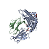
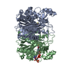
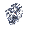
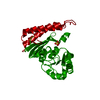
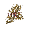
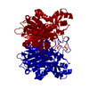
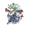
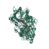
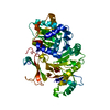
| Title | Crystal Structure of Hendra Virus Attachment G Glycoprotein in Complex with Receptor Ephrin-B2 | ||||||||||||
 Components Components |
| ||||||||||||
 Keywords Keywords |  VIRAL PROTEIN/SIGNALING PROTEIN / attachment / glycoprotein / G protein / VIRAL PROTEIN / receptor / VIRAL PROTEIN-SIGNALING PROTEIN complex VIRAL PROTEIN/SIGNALING PROTEIN / attachment / glycoprotein / G protein / VIRAL PROTEIN / receptor / VIRAL PROTEIN-SIGNALING PROTEIN complex | ||||||||||||
| Function / homology |  Function and homology information Function and homology informationvenous blood vessel morphogenesis / nephric duct morphogenesis / positive regulation of aorta morphogenesis / positive regulation of cardiac muscle cell differentiation / exo-alpha-sialidase activity / lymph vessel development / regulation of chemotaxis / cell migration involved in sprouting angiogenesis / regulation of postsynaptic neurotransmitter receptor internalization / adherens junction organization ...venous blood vessel morphogenesis / nephric duct morphogenesis / positive regulation of aorta morphogenesis / positive regulation of cardiac muscle cell differentiation / exo-alpha-sialidase activity / lymph vessel development / regulation of chemotaxis / cell migration involved in sprouting angiogenesis / regulation of postsynaptic neurotransmitter receptor internalization / adherens junction organization / EPH-Ephrin signaling / Ephrin signaling / blood vessel morphogenesis / regulation of postsynaptic membrane neurotransmitter receptor levels / keratinocyte proliferation / EPH-ephrin mediated repulsion of cells / anatomical structure morphogenesis / ephrin receptor signaling pathway / negative regulation of keratinocyte proliferation / EPHB-mediated forward signaling / T cell costimulation / ephrin receptor binding / axon guidance / postsynaptic density membrane / animal organ morphogenesis / adherens junction / Schaffer collateral - CA1 synapse / negative regulation of neuron projection development / virus receptor activity / cell-cell signaling / host cell surface / host cell surface receptor binding / cell adhesion / symbiont entry into host cell / focal adhesion / viral envelope / glutamatergic synapse / positive regulation of cell population proliferation / virion attachment to host cell / host cell plasma membrane / virion membrane / membrane / plasma membraneSimilarity search - Function | ||||||||||||
| Biological species |  Hendra henipavirus Hendra henipavirus Homo sapiens (human) Homo sapiens (human) | ||||||||||||
| Method | X-RAY DIFFRACTION / SYNCHROTRON / MOLECULAR REPLACEMENT / Resolution: 2.7 Å | ||||||||||||
 Authors Authors | Xu, K. / Nikolov, D.B. | ||||||||||||
| Funding support |  United States, 3items United States, 3items
| ||||||||||||
 Citation Citation | Journal: PLoS ONE / Year: 2012 Title: New insights into the Hendra virus attachment and entry process from structures of the virus G glycoprotein and its complex with Ephrin-B2. Authors: Xu, K. / Chan, Y.P. / Rajashankar, K.R. / Khetawat, D. / Yan, L. / Kolev, M.V. / Broder, C.C. / Nikolov, D.B. #1: Journal: Glycobiology / Year: 2012 Title: Site occupancy and glycan compositional analysis of two soluble recombinant forms of the attachment glycoprotein of Hendra virus. Authors: Colgrave, M.L. / Snelling, H.J. / Shiell, B.J. / Feng, Y.R. / Chan, Y.P. / Bossart, K.N. / Xu, K. / Nikolov, D.B. / Broder, C.C. / Michalski, W.P. | ||||||||||||
| History |
|
- Structure visualization
| Structure viewer | Molecule: MolmilJmol/JSmol |
|---|
- Downloads & links
-Download
| PDBx/mmCIF format | 6pdl.cif.gz | 469.6 KB | Display | PDBx/mmCIF format |
|---|---|---|---|---|
| PDB format | pdb6pdl.ent.gz | 385.5 KB | Display | PDB format |
| PDBx/mmJSON format | 6pdl.json.gz | Tree view | PDBx/mmJSON format | |
| Others |  Other downloads Other downloads |
-Validation report
| Arichive directory | https://data.pdbj.org/pub/pdb/validation_reports/pd/6pdlftp://data.pdbj.org/pub/pdb/validation_reports/pd/6pdl | HTTPS FTP |
|---|
-Related structure data
| Related structure data | 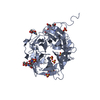 6pd4C 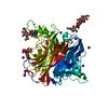 3d11S S: Starting model for refinement C: citing same article ( |
|---|---|
| Similar structure data |

-Links
 PDBj
PDBj


- Assembly
| Deposited unit | 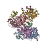
| |||||||||||||||||||||||||||||||||||||||||||||||||||||||||||||||||||||||||||||||||||||||||||
|---|---|---|---|---|---|---|---|---|---|---|---|---|---|---|---|---|---|---|---|---|---|---|---|---|---|---|---|---|---|---|---|---|---|---|---|---|---|---|---|---|---|---|---|---|---|---|---|---|---|---|---|---|---|---|---|---|---|---|---|---|---|---|---|---|---|---|---|---|---|---|---|---|---|---|---|---|---|---|---|---|---|---|---|---|---|---|---|---|---|---|---|---|
| 1 | 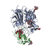
| |||||||||||||||||||||||||||||||||||||||||||||||||||||||||||||||||||||||||||||||||||||||||||
| 2 | 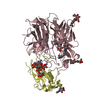
| |||||||||||||||||||||||||||||||||||||||||||||||||||||||||||||||||||||||||||||||||||||||||||
| 3 | 
| |||||||||||||||||||||||||||||||||||||||||||||||||||||||||||||||||||||||||||||||||||||||||||
| 4 | 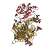
| |||||||||||||||||||||||||||||||||||||||||||||||||||||||||||||||||||||||||||||||||||||||||||
| Unit cell |
| |||||||||||||||||||||||||||||||||||||||||||||||||||||||||||||||||||||||||||||||||||||||||||
| Noncrystallographic symmetry (NCS) | NCS domain:
NCS domain segments:
|