 Movie
Movie Controller

[English] 日本語
 Yorodumi
- PDB-2vsm: Nipah virus attachment glycoprotein in complex with human cell su... -
+ Open data

- Basic information
| Entry | Database: PDB / ID: 2vsm | ||||||
|---|---|---|---|---|---|---|---|
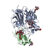
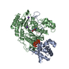
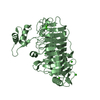
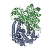
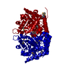
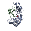
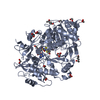
| Title | Nipah virus attachment glycoprotein in complex with human cell surface receptor ephrinB2 | ||||||
 Components Components |
| ||||||
 Keywords Keywords |  HYDROLASE / DEVELOPMENTAL PROTEIN / HENIPAVIRUS / NEUROGENESIS / GLYCOPROTEIN / PARAMYXOVIRUS / ENVELOPE PROTEIN / CELL SURFACE RECEPTOR / HENDRA / VIRION / EPHRIN / COMPLEX / MEMBRANE / B2 / EFN / NIV / EPH / HEV / HEV-G / NIPAH / VIRUS / NIV-G / PHOSPHOPROTEIN / DIFFERENTIATION / VIRAL ATTACHMENT / SIGNAL-ANCHOR / HEMAGGLUTININ / TRANSMEMBRANE HYDROLASE / DEVELOPMENTAL PROTEIN / HENIPAVIRUS / NEUROGENESIS / GLYCOPROTEIN / PARAMYXOVIRUS / ENVELOPE PROTEIN / CELL SURFACE RECEPTOR / HENDRA / VIRION / EPHRIN / COMPLEX / MEMBRANE / B2 / EFN / NIV / EPH / HEV / HEV-G / NIPAH / VIRUS / NIV-G / PHOSPHOPROTEIN / DIFFERENTIATION / VIRAL ATTACHMENT / SIGNAL-ANCHOR / HEMAGGLUTININ / TRANSMEMBRANE | ||||||
| Function / homology |  Function and homology information Function and homology informationvenous blood vessel morphogenesis / nephric duct morphogenesis / positive regulation of aorta morphogenesis / positive regulation of cardiac muscle cell differentiation / membrane fusion involved in viral entry into host cell / exo-alpha-sialidase activity / lymph vessel development / regulation of chemotaxis / cell migration involved in sprouting angiogenesis / regulation of postsynaptic neurotransmitter receptor internalization ...venous blood vessel morphogenesis / nephric duct morphogenesis / positive regulation of aorta morphogenesis / positive regulation of cardiac muscle cell differentiation / membrane fusion involved in viral entry into host cell / exo-alpha-sialidase activity / lymph vessel development / regulation of chemotaxis / cell migration involved in sprouting angiogenesis / regulation of postsynaptic neurotransmitter receptor internalization / adherens junction organization / EPH-Ephrin signaling / Ephrin signaling / blood vessel morphogenesis / regulation of postsynaptic membrane neurotransmitter receptor levels / keratinocyte proliferation / EPH-ephrin mediated repulsion of cells / anatomical structure morphogenesis / negative regulation of keratinocyte proliferation / ephrin receptor signaling pathway / EPHB-mediated forward signaling / T cell costimulation / ephrin receptor binding / axon guidance / animal organ morphogenesis / postsynaptic density membrane / adherens junction / Schaffer collateral - CA1 synapse / virus receptor activity / cell-cell signaling / negative regulation of neuron projection development / presynaptic membrane / clathrin-dependent endocytosis of virus by host cell / cell adhesion / host cell surface receptor binding / focal adhesion / viral envelope / glutamatergic synapse / positive regulation of cell population proliferation / virion attachment to host cell / host cell plasma membrane / virion membrane / membrane / identical protein binding / plasma membraneSimilarity search - Function | ||||||
| Biological species |  Nipah virus Nipah virus Homo sapiens (human) Homo sapiens (human) | ||||||
| Method | X-RAY DIFFRACTION / SYNCHROTRON / MOLECULAR REPLACEMENT / Resolution: 1.8 Å | ||||||
 Authors Authors | Bowden, T.A. / Aricescu, A.R. / Gilbert, R.J. / Grimes, J.M. / Jones, E.Y. / Stuart, D.I. | ||||||
 Citation Citation | Journal: Nat.Struct.Mol.Biol. / Year: 2008 Title: Structural Basis of Nipah and Hendra Virus Attachment to Their Cell-Surface Receptor Ephrin-B2 Authors: Bowden, T.A. / Aricescu, A.R. / Gilbert, R.J. / Grimes, J.M. / Jones, E.Y. / Stuart, D.I. | ||||||
| History |
| ||||||
| Remark 700 | SHEET THE SHEET STRUCTURE OF THIS MOLECULE IS BIFURCATED. IN ORDER TO REPRESENT THIS FEATURE IN ... SHEET THE SHEET STRUCTURE OF THIS MOLECULE IS BIFURCATED. IN ORDER TO REPRESENT THIS FEATURE IN THE SHEET RECORDS BELOW, TWO SHEETS ARE DEFINED. |
- Structure visualization
| Structure viewer | Molecule: MolmilJmol/JSmol |
|---|
- Downloads & links
-Download
| PDBx/mmCIF format | 2vsm.cif.gz | 145.9 KB | Display | PDBx/mmCIF format |
|---|---|---|---|---|
| PDB format | pdb2vsm.ent.gz | 112.7 KB | Display | PDB format |
| PDBx/mmJSON format | 2vsm.json.gz | Tree view | PDBx/mmJSON format | |
| Others |  Other downloads Other downloads |
-Validation report
| Arichive directory | https://data.pdbj.org/pub/pdb/validation_reports/vs/2vsmftp://data.pdbj.org/pub/pdb/validation_reports/vs/2vsm | HTTPS FTP |
|---|
-Related structure data
| Related structure data |  2vskC  1nukS  1v3eS C: citing same article ( S: Starting model for refinement |
|---|---|
| Similar structure data |
-Links
 PDBj
PDBj


- Assembly
| Deposited unit | 
| ||||||||
|---|---|---|---|---|---|---|---|---|---|
| 1 |
| ||||||||
| Unit cell |
|
-Components
| #1: Protein | / NIV-G Mass: 46839.293 Da / Num. of mol.: 1 Fragment: B-PROPELLER, EPHRIN BINDING DOMAIN, RESIDUES 188-602 Source method: isolated from a genetically manipulated source Details: N-ACETYLGLUCOSAMINE LINKAGES OBSERVED IN / Source: (gene. exp.) Nipah virus / Description: SYNTHETICALLY OPTIMIZED CDNA (GENEART) / Plasmid: PHLSEC / Cell line (production host): HEK293T / Production host: Homo sapiens (human) / References: UniProt: Q9IH62, exo-alpha-sialidase | ||
|---|---|---|---|
| #2: Protein | Ephrin B2 / EFNB2 / EPH-RELATED RECEPTOR TYROSINE KINASE LIGAND 5 / LERK-5 / HTK LIGAND / HTK-L Mass: 16049.366 Da / Num. of mol.: 1 / Fragment: RECEPTOR-BINDING DOMAIN, RESIDUES 28-165 Source method: isolated from a genetically manipulated source Details: N-ACETYLGLUCOSAMINE LINKAGE OBSERVED IN / Source: (gene. exp.) Homo sapiens (human) / Plasmid: PHLSEC / Cell line (production host): HEK293T / Production host: Homo sapiens (human) / References: UniProt: P52799 | ||
| #3: Chemical | ChemComp-IPA / Isopropyl alcohol  Mass: 60.095 Da / Num. of mol.: 1 / Source method: obtained synthetically / Formula: C3H8O / Comment: alkaloid*YM Mass: 60.095 Da / Num. of mol.: 1 / Source method: obtained synthetically / Formula: C3H8O / Comment: alkaloid*YM | ||
| #4: Sugar | ChemComp-NAG / N-Acetylglucosamine  Type: D-saccharide, beta linking / Mass: 221.208 Da / Num. of mol.: 4 Type: D-saccharide, beta linking / Mass: 221.208 Da / Num. of mol.: 4Source method: isolated from a genetically manipulated source Formula: C8H15NO6 #5: Water | ChemComp-HOH / | Water Mass: 18.015 Da / Num. of mol.: 705 / Source method: isolated from a natural source / Formula: H2O Mass: 18.015 Da / Num. of mol.: 705 / Source method: isolated from a natural source / Formula: H2O |
-Experimental details
-Experiment
| Experiment | Method: X-RAY DIFFRACTION / Number of used crystals: 1 |
|---|
- Sample preparation
| Crystal | Density Matthews: 2.3 Å3/Da / Density % sol: 46 % / Description: NONE |
|---|---|
| Crystal grow | pH: 5.6 Details: 18% ISOPROPANOL, 18% PEG 3350 AND 0.1 M TRI-CITRATE BUFFER PH 5.6 |
-Data collection
| Diffraction | Mean temperature: 77.2 K |
|---|---|
| Diffraction source | Source: SYNCHROTRON / Site: ESRF  / Beamline: ID14-2 / Wavelength: 0.933 / Beamline: ID14-2 / Wavelength: 0.933 |
| Detector | Type: ADSC CCD / Detector: CCD / Date: Dec 17, 2006 / Details: MIRRORS |
| Radiation | Monochromator: SI(III) / Protocol: SINGLE WAVELENGTH / Monochromatic (M) / Laue (L): M / Scattering type: x-ray |
| Radiation wavelength | Wavelength: 0.933 Å / Relative weight: 1 |
| Reflection | Resolution: 1.8→30 Å / Num. obs: 55159 / % possible obs: 98.9 % / Observed criterion σ(I): -3 / Redundancy: 6.9 % / Rmerge(I) obs: 0.09 / Net I/σ(I): 15 |
| Reflection shell | Resolution: 1.8→1.86 Å / Redundancy: 5.5 % / Rmerge(I) obs: 0.52 / Mean I/σ(I) obs: 2.3 / % possible all: 90.4 |
- Processing
| Software |
| ||||||||||||||||||||||||||||||||||||||||||||||||||||||||||||||||||||||||||||||||||||||||||||||||||||||||||||||||||||||||||||||||||||||||||||||||||||||||||||||||||||||||||||||||||||||
|---|---|---|---|---|---|---|---|---|---|---|---|---|---|---|---|---|---|---|---|---|---|---|---|---|---|---|---|---|---|---|---|---|---|---|---|---|---|---|---|---|---|---|---|---|---|---|---|---|---|---|---|---|---|---|---|---|---|---|---|---|---|---|---|---|---|---|---|---|---|---|---|---|---|---|---|---|---|---|---|---|---|---|---|---|---|---|---|---|---|---|---|---|---|---|---|---|---|---|---|---|---|---|---|---|---|---|---|---|---|---|---|---|---|---|---|---|---|---|---|---|---|---|---|---|---|---|---|---|---|---|---|---|---|---|---|---|---|---|---|---|---|---|---|---|---|---|---|---|---|---|---|---|---|---|---|---|---|---|---|---|---|---|---|---|---|---|---|---|---|---|---|---|---|---|---|---|---|---|---|---|---|---|---|
| Refinement | Method to determine structure: MOLECULAR REPLACEMENT Starting model: PDB ENTRY 1NUK AND 1V3E Resolution: 1.8→30 Å / Cor.coef. Fo:Fc: 0.96 / Cor.coef. Fo:Fc free: 0.936 / SU B: 2.237 / SU ML: 0.071 / Cross valid method: THROUGHOUT / ESU R: 0.115 / ESU R Free: 0.115 / Stereochemistry target values: MAXIMUM LIKELIHOOD / Details: HYDROGENS HAVE BEEN ADDED IN THE RIDING POSITIONS.
| ||||||||||||||||||||||||||||||||||||||||||||||||||||||||||||||||||||||||||||||||||||||||||||||||||||||||||||||||||||||||||||||||||||||||||||||||||||||||||||||||||||||||||||||||||||||
| Solvent computation | Ion probe radii: 0.8 Å / Shrinkage radii: 0.8 Å / VDW probe radii: 1.4 Å / Solvent model: MASK | ||||||||||||||||||||||||||||||||||||||||||||||||||||||||||||||||||||||||||||||||||||||||||||||||||||||||||||||||||||||||||||||||||||||||||||||||||||||||||||||||||||||||||||||||||||||
| Displacement parameters | Biso mean: 11.79 Å2
| ||||||||||||||||||||||||||||||||||||||||||||||||||||||||||||||||||||||||||||||||||||||||||||||||||||||||||||||||||||||||||||||||||||||||||||||||||||||||||||||||||||||||||||||||||||||
| Refinement step | Cycle: LAST / Resolution: 1.8→30 Å
| ||||||||||||||||||||||||||||||||||||||||||||||||||||||||||||||||||||||||||||||||||||||||||||||||||||||||||||||||||||||||||||||||||||||||||||||||||||||||||||||||||||||||||||||||||||||
| Refine LS restraints |
|