 Movie
Movie Controller

[English] 日本語
 Yorodumi
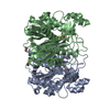
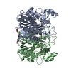
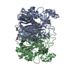
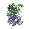
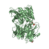
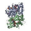
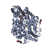
- PDB-3t6s: Crystal structure of CerJ from Streptomyces tendae in Complex with CoA -
+ Open data

- Basic information
| Entry | Database: PDB / ID: 3t6s | ||||||
|---|---|---|---|---|---|---|---|
| Title | Crystal structure of CerJ from Streptomyces tendae in Complex with CoA | ||||||
 Components Components | (CerJ) x 2 | ||||||
 Keywords Keywords |  TRANSFERASE / Thiloase superfamily / FabH-like fold / Acyltransferase / O-malonyl transferase TRANSFERASE / Thiloase superfamily / FabH-like fold / Acyltransferase / O-malonyl transferase | ||||||
| Function / homology |  Function and homology information Function and homology information3-oxoacyl-[acyl-carrier-protein] synthase activity / fatty acid biosynthetic process Similarity search - Function | ||||||
| Biological species |  Streptomyces tendae (bacteria) Streptomyces tendae (bacteria) | ||||||
| Method | X-RAY DIFFRACTION / SYNCHROTRON / Resolution: 2 Å | ||||||
 Authors Authors | Zocher, G. / Bretschneider, T. / Hertweck, C. / Stehle, T. | ||||||
 Citation Citation | Journal: Nat.Chem.Biol. / Year: 2011 Title: A ketosynthase homolog uses malonyl units to form esters in cervimycin biosynthesis. Authors: Bretschneider, T. / Zocher, G. / Unger, M. / Scherlach, K. / Stehle, T. / Hertweck, C. | ||||||
| History |
|
- Structure visualization
| Structure viewer | Molecule: MolmilJmol/JSmol |
|---|
- Downloads & links
-Download
| PDBx/mmCIF format | 3t6s.cif.gz | 277.1 KB | Display | PDBx/mmCIF format |
|---|---|---|---|---|
| PDB format | pdb3t6s.ent.gz | 235 KB | Display | PDB format |
| PDBx/mmJSON format | 3t6s.json.gz | Tree view | PDBx/mmJSON format | |
| Others |  Other downloads Other downloads |
-Validation report
| Arichive directory | https://data.pdbj.org/pub/pdb/validation_reports/t6/3t6sftp://data.pdbj.org/pub/pdb/validation_reports/t6/3t6s | HTTPS FTP |
|---|
-Related structure data


-Links
 PDBj
PDBj


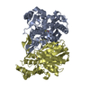
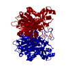
- Assembly
| Deposited unit | 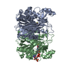
| ||||||||
|---|---|---|---|---|---|---|---|---|---|
| 1 |
| ||||||||
| Unit cell |
| ||||||||
| Components on special symmetry positions |
|
-Components
| #1: Protein | Mass: 37604.203 Da / Num. of mol.: 1 Source method: isolated from a genetically manipulated source Source: (gene. exp.) Streptomyces tendae (bacteria) / Production host: Escherichia coli (E. coli) / References: UniProt: H2L2M1*PLUS |
|---|---|
| #2: Protein | Mass: 37572.203 Da / Num. of mol.: 1 Source method: isolated from a genetically manipulated source Source: (gene. exp.) Streptomyces tendae (bacteria) / Production host: Escherichia coli (E. coli) / References: UniProt: H2L2M1*PLUS |
| #3: Chemical | ChemComp-COA / Coenzyme A  Mass: 767.534 Da / Num. of mol.: 1 / Source method: obtained synthetically / Formula: C21H36N7O16P3S Mass: 767.534 Da / Num. of mol.: 1 / Source method: obtained synthetically / Formula: C21H36N7O16P3S |
| #4: Water | ChemComp-HOH / Water Mass: 18.015 Da / Num. of mol.: 410 / Source method: isolated from a natural source / Formula: H2O Mass: 18.015 Da / Num. of mol.: 410 / Source method: isolated from a natural source / Formula: H2O |
-Experimental details
-Experiment
| Experiment | Method: X-RAY DIFFRACTION / Number of used crystals: 1 |
|---|
- Sample preparation
| Crystal | Density Matthews: 2.32 Å3/Da / Density % sol: 46.97 % |
|---|---|
| Crystal grow | Temperature: 293 K / Method: vapor diffusion, hanging drop / pH: 8.5 Details: 22.5% PEG 4000, 100 mM Tris/HCl pH 8.5, 200 mM sodium acetate, VAPOR DIFFUSION, HANGING DROP, temperature 293K |
-Data collection
| Diffraction | Mean temperature: 100 K |
|---|---|
| Diffraction source | Source: SYNCHROTRON / Site: SLS  / Beamline: X06DA / Wavelength: 1 Å / Beamline: X06DA / Wavelength: 1 Å |
| Detector | Type: MARMOSAIC 225 mm CCD / Detector: CCD / Date: Sep 27, 2010 |
| Radiation | Monochromator: DCCM / Protocol: SINGLE WAVELENGTH / Monochromatic (M) / Laue (L): M / Scattering type: x-ray |
| Radiation wavelength | Wavelength: 1 Å / Relative weight: 1 |
| Reflection | Resolution: 2→30 Å / Num. all: 48366 / Num. obs: 48321 / % possible obs: 99.9 % / Observed criterion σ(F): 3 / Observed criterion σ(I): 3 |
| Reflection shell | Resolution: 2→2.05 Å / Rmerge(I) obs: 0.491 / Mean I/σ(I) obs: 3.73 / Num. unique all: 3522 / % possible all: 100 |
- Processing
| Software |
| ||||||||||||||||||||||||||||||||||||||||||||||||||||||||||||||||||||||||||||||||||||||||||||||||||||||||||||||||||||||||||||||||||||||||||||||||||||||||||||||||||||||||||
|---|---|---|---|---|---|---|---|---|---|---|---|---|---|---|---|---|---|---|---|---|---|---|---|---|---|---|---|---|---|---|---|---|---|---|---|---|---|---|---|---|---|---|---|---|---|---|---|---|---|---|---|---|---|---|---|---|---|---|---|---|---|---|---|---|---|---|---|---|---|---|---|---|---|---|---|---|---|---|---|---|---|---|---|---|---|---|---|---|---|---|---|---|---|---|---|---|---|---|---|---|---|---|---|---|---|---|---|---|---|---|---|---|---|---|---|---|---|---|---|---|---|---|---|---|---|---|---|---|---|---|---|---|---|---|---|---|---|---|---|---|---|---|---|---|---|---|---|---|---|---|---|---|---|---|---|---|---|---|---|---|---|---|---|---|---|---|---|---|---|---|---|
| Refinement | Resolution: 2→29.65 Å / Cor.coef. Fo:Fc: 0.97 / Cor.coef. Fo:Fc free: 0.957 / SU B: 8.213 / SU ML: 0.101 / Cross valid method: THROUGHOUT / ESU R: 0.157 / ESU R Free: 0.139 / Stereochemistry target values: MAXIMUM LIKELIHOOD / Details: HYDROGENS HAVE BEEN ADDED IN THE RIDING POSITIONS
| ||||||||||||||||||||||||||||||||||||||||||||||||||||||||||||||||||||||||||||||||||||||||||||||||||||||||||||||||||||||||||||||||||||||||||||||||||||||||||||||||||||||||||
| Solvent computation | Ion probe radii: 0.8 Å / Shrinkage radii: 0.8 Å / VDW probe radii: 1.4 Å / Solvent model: BABINET MODEL WITH MASK | ||||||||||||||||||||||||||||||||||||||||||||||||||||||||||||||||||||||||||||||||||||||||||||||||||||||||||||||||||||||||||||||||||||||||||||||||||||||||||||||||||||||||||
| Displacement parameters | Biso mean: 46.835 Å2
| ||||||||||||||||||||||||||||||||||||||||||||||||||||||||||||||||||||||||||||||||||||||||||||||||||||||||||||||||||||||||||||||||||||||||||||||||||||||||||||||||||||||||||
| Refinement step | Cycle: LAST / Resolution: 2→29.65 Å
| ||||||||||||||||||||||||||||||||||||||||||||||||||||||||||||||||||||||||||||||||||||||||||||||||||||||||||||||||||||||||||||||||||||||||||||||||||||||||||||||||||||||||||
| Refine LS restraints |
| ||||||||||||||||||||||||||||||||||||||||||||||||||||||||||||||||||||||||||||||||||||||||||||||||||||||||||||||||||||||||||||||||||||||||||||||||||||||||||||||||||||||||||
| LS refinement shell | Resolution: 2→2.051 Å / Total num. of bins used: 20
| ||||||||||||||||||||||||||||||||||||||||||||||||||||||||||||||||||||||||||||||||||||||||||||||||||||||||||||||||||||||||||||||||||||||||||||||||||||||||||||||||||||||||||
| Refinement TLS params. | Method: refined / Refine-ID: X-RAY DIFFRACTION
| ||||||||||||||||||||||||||||||||||||||||||||||||||||||||||||||||||||||||||||||||||||||||||||||||||||||||||||||||||||||||||||||||||||||||||||||||||||||||||||||||||||||||||
| Refinement TLS group |
|
