 Movie
Movie Controller

[English] 日本語
 Yorodumi
- PDB-6nxd: TYPE I L-ASPARAGINASE FROM ESCHERICHIA COLI IN COMPLEX WITH CITRA... -
+ Open data

- Basic information
| Entry | Database: PDB / ID: 6nxd | ||||||
|---|---|---|---|---|---|---|---|
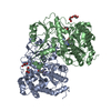
| Title | TYPE I L-ASPARAGINASE FROM ESCHERICHIA COLI IN COMPLEX WITH CITRATE AT PH 4 | ||||||
 Components Components | L-asparaginase 1 Asparaginase Asparaginase | ||||||
 Keywords Keywords | HYDROLASE / hydrolysis of L-asparagine | ||||||
| Function / homology |  Function and homology information Function and homology informationasparagine catabolic process via L-aspartate / asparaginase / asparaginase activity / protein homotetramerization / identical protein binding / cytosol / cytoplasmSimilarity search - Function | ||||||
| Biological species |  Escherichia coli (E. coli) Escherichia coli (E. coli) | ||||||
| Method | X-RAY DIFFRACTION / SYNCHROTRON / Resolution: 1.9 Å | ||||||
 Authors Authors | Lubkowski, J. / Wlodawer, A. | ||||||
 Citation Citation | Journal: J. Mol. Biol. / Year: 2007 Title: Crystal structure and allosteric regulation of the cytoplasmic Escherichia coli L-asparaginase I. Authors: Yun, M.K. / Nourse, A. / White, S.W. / Rock, C.O. / Heath, R.J. | ||||||
| History |
| ||||||
| Remark 0 | THIS ENTRY 1ZET REFLECTS AN ALTERNATIVE MODELING OF THE ORIGINAL DATA IN 2P2N, DETERMINED BY YUN, M. ...THIS ENTRY 1ZET REFLECTS AN ALTERNATIVE MODELING OF THE ORIGINAL DATA IN 2P2N, DETERMINED BY YUN, M.K., NOURSE, A., WHITE, S.W., ROCK, C.O., HEATH, R.J. |
- Structure visualization
| Structure viewer | Molecule: MolmilJmol/JSmol |
|---|
- Downloads & links
-Download
| PDBx/mmCIF format | 6nxd.cif.gz | 264 KB | Display | PDBx/mmCIF format |
|---|---|---|---|---|
| PDB format | pdb6nxd.ent.gz | 217.4 KB | Display | PDB format |
| PDBx/mmJSON format | 6nxd.json.gz | Tree view | PDBx/mmJSON format | |
| Others |  Other downloads Other downloads |
-Validation report
| Arichive directory | https://data.pdbj.org/pub/pdb/validation_reports/nx/6nxdftp://data.pdbj.org/pub/pdb/validation_reports/nx/6nxd | HTTPS FTP |
|---|
-Related structure data
| Related structure data |  6nx6C  6nx7C  6nx8C  6nx9C  6nxaC  6nxbC  6nxcC C: citing same article ( |
|---|---|
| Similar structure data |

-Links
 PDBj
PDBj- Assembly
| Deposited unit | 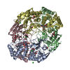
| ||||||||
|---|---|---|---|---|---|---|---|---|---|
| 1 |
| ||||||||
| Unit cell |
|
-Components
-Protein , 1 types, 4 molecules ABCD
| #1: Protein | Asparaginase / L-asparaginase I / L-ASNase I / L-asparagine amidohydrolase I Mass: 39335.461 Da / Num. of mol.: 4 Source method: isolated from a genetically manipulated source Source: (gene. exp.) Escherichia coli (strain K12) (bacteria)Strain: K12 / Gene: ansA, b1767, JW1756 / Plasmid: pET-15b / Cell (production host): mesophilic bacteria / Production host: Escherichia coli BL21(DE3) (bacteria) / Strain (production host): BL21(DE3) / References: UniProt: P0A962, asparaginase |
|---|
-Non-polymers , 5 types, 595 molecules 


| #2: Chemical | ChemComp-CL / Chloride Mass: 35.453 Da / Num. of mol.: 10 / Source method: obtained synthetically / Formula: Cl Mass: 35.453 Da / Num. of mol.: 10 / Source method: obtained synthetically / Formula: Cl#3: Chemical | ChemComp-CIT / Citric acid Mass: 192.124 Da / Num. of mol.: 4 / Source method: obtained synthetically / Formula: C6H8O7 Mass: 192.124 Da / Num. of mol.: 4 / Source method: obtained synthetically / Formula: C6H8O7#4: Chemical | ChemComp-ASN / Asparagine Type: L-peptide linking / Mass: 132.118 Da / Num. of mol.: 4 / Source method: obtained synthetically / Formula: C4H8N2O3 Type: L-peptide linking / Mass: 132.118 Da / Num. of mol.: 4 / Source method: obtained synthetically / Formula: C4H8N2O3#5: Chemical | ChemComp-EDO / Ethylene glycol Mass: 62.068 Da / Num. of mol.: 13 / Source method: obtained synthetically / Formula: C2H6O2 Mass: 62.068 Da / Num. of mol.: 13 / Source method: obtained synthetically / Formula: C2H6O2#6: Water | ChemComp-HOH / | WaterMass: 18.015 Da / Num. of mol.: 564 / Source method: isolated from a natural source / Formula: H2O |
|---|
-Experimental details
-Experiment
| Experiment | Method: X-RAY DIFFRACTION / Number of used crystals: 1 |
|---|
- Sample preparation
| Crystal | Density Matthews: 2.14 Å3/Da / Density % sol: 42.41 % / Description: AUTHOR USED THE SF DATA FROM ENTRY 2P2N. |
|---|---|
| Crystal grow | Temperature: 291 K / Method: vapor diffusion, hanging drop / pH: 4 / Details: citric acid, sodium chloride, pH 4.0 |
-Data collection
| Diffraction | Mean temperature: 100 K / Serial crystal experiment: N |
|---|---|
| Diffraction source | Source: SYNCHROTRON / Site: APS  / Beamline: 22-ID / Wavelength: 1.01259 Å / Beamline: 22-ID / Wavelength: 1.01259 Å |
| Detector | Type: MARRESEARCH / Detector: CCD / Date: Mar 6, 2005 |
| Radiation | Protocol: SINGLE WAVELENGTH / Monochromatic (M) / Laue (L): M / Scattering type: x-ray |
| Radiation wavelength | Wavelength: 1.01259 Å / Relative weight: 1 |
| Reflection | Resolution: 1.9→50 Å / Num. obs: 95017 / % possible obs: 90.6 % / Observed criterion σ(I): -3 / Redundancy: 3.6 % / Net I/σ(I): 17.9 |
| Reflection shell | Resolution: 1.9→1.97 Å |
- Processing
| Software |
| ||||||||||||||||||||||||||||||||||||||||||||||||||||||||||||
|---|---|---|---|---|---|---|---|---|---|---|---|---|---|---|---|---|---|---|---|---|---|---|---|---|---|---|---|---|---|---|---|---|---|---|---|---|---|---|---|---|---|---|---|---|---|---|---|---|---|---|---|---|---|---|---|---|---|---|---|---|---|
| Refinement | Resolution: 1.9→45.17 Å / Cor.coef. Fo:Fc: 0.936 / Cor.coef. Fo:Fc free: 0.913 / SU B: 3.663 / SU ML: 0.109 / SU R Cruickshank DPI: 0.1885 / Cross valid method: THROUGHOUT / σ(F): 0 / ESU R: 0.189 / ESU R Free: 0.166 Details: HYDROGENS HAVE BEEN ADDED IN THE RIDING POSITIONS U VALUES : REFINED INDIVIDUALLY
| ||||||||||||||||||||||||||||||||||||||||||||||||||||||||||||
| Solvent computation | Ion probe radii: 0.8 Å / Shrinkage radii: 0.8 Å / VDW probe radii: 1.2 Å | ||||||||||||||||||||||||||||||||||||||||||||||||||||||||||||
| Displacement parameters | Biso max: 103.22 Å2 / Biso mean: 21.296 Å2 / Biso min: 6.53 Å2
| ||||||||||||||||||||||||||||||||||||||||||||||||||||||||||||
| Refinement step | Cycle: final / Resolution: 1.9→45.17 Å
| ||||||||||||||||||||||||||||||||||||||||||||||||||||||||||||
| Refine LS restraints |
| ||||||||||||||||||||||||||||||||||||||||||||||||||||||||||||
| LS refinement shell | Resolution: 1.9→1.949 Å / Rfactor Rfree error: 0 / Total num. of bins used: 20
|
