 Movie
Movie Controller

[English] 日本語
 Yorodumi
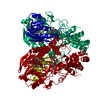
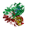
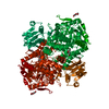
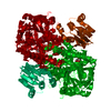
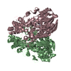
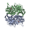
- PDB-2cfu: Crystal structure of SdsA1, an alkylsulfatase from Pseudomonas ae... -
+ Open data

- Basic information
| Entry | Database: PDB / ID: 2cfu | ||||||
|---|---|---|---|---|---|---|---|
| Title | Crystal structure of SdsA1, an alkylsulfatase from Pseudomonas aeruginosa, in complex with 1-decane-sulfonic-acid. | ||||||
 Components Components | SDSA1 | ||||||
 Keywords Keywords |  HYDROLASE / SDS-HYDROLASE / SDSA1 / LACTAMASE HYDROLASE / SDS-HYDROLASE / SDSA1 / LACTAMASE | ||||||
| Function / homology |  Function and homology information Function and homology informationlinear primary-alkylsulfatase / linear primary-alkylsulfatase activity / dodecyl sulfate metabolic process / outer membrane-bounded periplasmic space / protein dimerization activity / identical protein binding / metal ion bindingSimilarity search - Function | ||||||
| Biological species |   PSEUDOMONAS AERUGINOSA (bacteria) PSEUDOMONAS AERUGINOSA (bacteria) | ||||||
| Method | X-RAY DIFFRACTION / SYNCHROTRON / MOLECULAR REPLACEMENT / Resolution: 1.9 Å | ||||||
 Authors Authors | Hagelueken, G. / Adams, T.M. / Wiehlmann, L. / Widow, U. / Kolmar, H. / Tuemmler, B. / Heinz, D.W. / Schubert, W.-D. | ||||||
 Citation Citation | Journal: Proc.Natl.Acad.Sci.USA / Year: 2006 Title: The Crystal Structure of Sdsa1, an Alkylsulfatase from Pseudomonas Aeruginosa, Defines a Third Class of Sulfatases. Authors: Hagelueken, G. / Adams, T.M. / Wiehlmann, L. / Widow, U. / Kolmar, H. / Tummler, B. / Heinz, D.W. / Schubert, W.-D. | ||||||
| History |
|
- Structure visualization
| Structure viewer | Molecule: MolmilJmol/JSmol |
|---|
- Downloads & links
-Download
| PDBx/mmCIF format | 2cfu.cif.gz | 156.5 KB | Display | PDBx/mmCIF format |
|---|---|---|---|---|
| PDB format | pdb2cfu.ent.gz | 123.8 KB | Display | PDB format |
| PDBx/mmJSON format | 2cfu.json.gz | Tree view | PDBx/mmJSON format | |
| Others |  Other downloads Other downloads |
-Validation report
| Arichive directory | https://data.pdbj.org/pub/pdb/validation_reports/cf/2cfuftp://data.pdbj.org/pub/pdb/validation_reports/cf/2cfu | HTTPS FTP |
|---|
-Related structure data
| Related structure data |  2cfzC  2cg2C  2cg3SC C: citing same article ( S: Starting model for refinement |
|---|---|
| Similar structure data |
-Links
 PDBj
PDBj


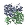
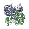
- Assembly
| Deposited unit | 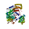
| ||||||||
|---|---|---|---|---|---|---|---|---|---|
| 1 | 
| ||||||||
| Unit cell |
|
-Components
-Protein , 1 types, 1 molecules A
| #1: Protein | Mass: 72673.031 Da / Num. of mol.: 1 Source method: isolated from a genetically manipulated source Source: (gene. exp.) PSEUDOMONAS AERUGINOSA (bacteria) / Strain: PAO1 / Production host: ESCHERICHIA COLI (E. coli) / References: UniProt: Q9I5I9 |
|---|
-Non-polymers , 5 types, 549 molecules 


| #2: Chemical | ChemComp-1DB /  Mass: 222.345 Da / Num. of mol.: 1 / Source method: obtained synthetically / Formula: C10H22O3S Mass: 222.345 Da / Num. of mol.: 1 / Source method: obtained synthetically / Formula: C10H22O3S | ||||||
|---|---|---|---|---|---|---|---|
| #3: Chemical | ChemComp-PEG / Diethylene glycol Mass: 106.120 Da / Num. of mol.: 8 / Source method: obtained synthetically / Formula: C4H10O3 Mass: 106.120 Da / Num. of mol.: 8 / Source method: obtained synthetically / Formula: C4H10O3#4: Chemical | ChemComp-IPA / | Isopropyl alcohol Mass: 60.095 Da / Num. of mol.: 1 / Source method: obtained synthetically / Formula: C3H8O / Comment: alkaloid*YM Mass: 60.095 Da / Num. of mol.: 1 / Source method: obtained synthetically / Formula: C3H8O / Comment: alkaloid*YM#5: Chemical |  Mass: 65.409 Da / Num. of mol.: 2 / Source method: obtained synthetically / Formula: Zn Mass: 65.409 Da / Num. of mol.: 2 / Source method: obtained synthetically / Formula: Zn#6: Water | ChemComp-HOH / | WaterMass: 18.015 Da / Num. of mol.: 537 / Source method: isolated from a natural source / Formula: H2O |
-Experimental details
-Experiment
| Experiment | Method: X-RAY DIFFRACTION / Number of used crystals: 1 |
|---|
- Sample preparation
| Crystal | Density Matthews: 2.6 Å3/Da / Density % sol: 52.7 % |
|---|---|
| Crystal grow | pH: 6 Details: 12% PEG 4000,10 % ISO-PROPANOL, 200 MM LICL,100 MM CITRATE PH6, pH 6.00 |
-Data collection
| Diffraction | Mean temperature: 100 K |
|---|---|
| Diffraction source | Source: SYNCHROTRON / Site: SLS  / Beamline: X06SA / Wavelength: 0.9001 / Beamline: X06SA / Wavelength: 0.9001 |
| Detector | Type: MARRESEARCH / Detector: CCD / Date: Jun 20, 2005 |
| Radiation | Protocol: SINGLE WAVELENGTH / Monochromatic (M) / Laue (L): M / Scattering type: x-ray |
| Radiation wavelength | Wavelength: 0.9001 Å / Relative weight: 1 |
| Reflection | Resolution: 1.9→50 Å / Num. obs: 68233 / % possible obs: 98 % / Observed criterion σ(I): 3 / Redundancy: 6.8 % / Rmerge(I) obs: 0.12 / Net I/σ(I): 12 |
| Reflection shell | Resolution: 1.9→1.95 Å / Redundancy: 7 % / Rmerge(I) obs: 0.52 / Mean I/σ(I) obs: 3.1 / % possible all: 98 |
- Processing
| Software |
| ||||||||||||||||||||||||||||||||||||||||||||||||||||||||||||||||||||||||||||||||||||||||||||||||||||||||||||||||||||||||||||||||||||||||||||||||||||||||||||||||||||||||||||||||||||||
|---|---|---|---|---|---|---|---|---|---|---|---|---|---|---|---|---|---|---|---|---|---|---|---|---|---|---|---|---|---|---|---|---|---|---|---|---|---|---|---|---|---|---|---|---|---|---|---|---|---|---|---|---|---|---|---|---|---|---|---|---|---|---|---|---|---|---|---|---|---|---|---|---|---|---|---|---|---|---|---|---|---|---|---|---|---|---|---|---|---|---|---|---|---|---|---|---|---|---|---|---|---|---|---|---|---|---|---|---|---|---|---|---|---|---|---|---|---|---|---|---|---|---|---|---|---|---|---|---|---|---|---|---|---|---|---|---|---|---|---|---|---|---|---|---|---|---|---|---|---|---|---|---|---|---|---|---|---|---|---|---|---|---|---|---|---|---|---|---|---|---|---|---|---|---|---|---|---|---|---|---|---|---|---|
| Refinement | Method to determine structure: MOLECULAR REPLACEMENT Starting model: PDB ENTRY 2CG3 Resolution: 1.9→74.54 Å / Cor.coef. Fo:Fc: 0.957 / Cor.coef. Fo:Fc free: 0.935 / SU B: 5.701 / SU ML: 0.09 / TLS residual ADP flag: LIKELY RESIDUAL / Cross valid method: THROUGHOUT / ESU R: 0.132 / ESU R Free: 0.131 / Stereochemistry target values: MAXIMUM LIKELIHOOD Details: HYDROGENS HAVE BEEN ADDED IN THE RIDING POSITIONS. ATOM RECORD CONTAINS RESIDUAL B FACTORS ONLY. RESIDUES 206-208 ARE DISORDERED AND WERE MODELLED BY WATER. CHAIN B 1-6.
| ||||||||||||||||||||||||||||||||||||||||||||||||||||||||||||||||||||||||||||||||||||||||||||||||||||||||||||||||||||||||||||||||||||||||||||||||||||||||||||||||||||||||||||||||||||||
| Solvent computation | Ion probe radii: 0.8 Å / Shrinkage radii: 0.8 Å / VDW probe radii: 1.4 Å / Solvent model: BABINET MODEL WITH MASK | ||||||||||||||||||||||||||||||||||||||||||||||||||||||||||||||||||||||||||||||||||||||||||||||||||||||||||||||||||||||||||||||||||||||||||||||||||||||||||||||||||||||||||||||||||||||
| Displacement parameters | Biso mean: 19.91 Å2
| ||||||||||||||||||||||||||||||||||||||||||||||||||||||||||||||||||||||||||||||||||||||||||||||||||||||||||||||||||||||||||||||||||||||||||||||||||||||||||||||||||||||||||||||||||||||
| Refinement step | Cycle: LAST / Resolution: 1.9→74.54 Å
| ||||||||||||||||||||||||||||||||||||||||||||||||||||||||||||||||||||||||||||||||||||||||||||||||||||||||||||||||||||||||||||||||||||||||||||||||||||||||||||||||||||||||||||||||||||||
| Refine LS restraints |
|