 Movie
Movie Controller


+ Open data

- Basic information
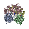
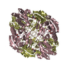
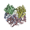
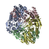
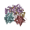
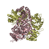
| Entry | Database: PDB / ID: 6nxa | ||||||
|---|---|---|---|---|---|---|---|
| Title | ECAII(D90T,K162T) MUTANT AT PH 7 | ||||||
 Components Components | L-asparaginase 2 Asparaginase Asparaginase | ||||||
 Keywords Keywords | HYDROLASE / inactive mutant / hydrolysis of L-asparagine | ||||||
| Function / homology |  Function and homology information Function and homology informationasparagine catabolic process / asparaginase / asparaginase activity / outer membrane-bounded periplasmic space / protein homotetramerization / periplasmic space / protein-containing complex / identical protein bindingSimilarity search - Function | ||||||
| Biological species |  Escherichia coli (E. coli) Escherichia coli (E. coli) | ||||||
| Method | X-RAY DIFFRACTION / FOURIER SYNTHESIS / Resolution: 1.93 Å | ||||||
 Authors Authors | Lubkowski, J. / Wlodawer, A. | ||||||
 Citation Citation | Journal: Sci Rep / Year: 2019 Title: Opportunistic complexes of E. coli L-asparaginases with citrate anions. Authors: Lubkowski, J. / Chan, W. / Wlodawer, A. #1: Journal: J. Mol. Biol. / Year: 2007Title: Crystal structure and allosteric regulation of the cytoplasmic Escherichia coli L-asparaginase I. Authors: Yun, M.K. / Nourse, A. / White, S.W. / Rock, C.O. / Heath, R.J. | ||||||
| History |
|
- Structure visualization
| Structure viewer | Molecule: MolmilJmol/JSmol |
|---|
- Downloads & links
-Download
| PDBx/mmCIF format | 6nxa.cif.gz | 267.9 KB | Display | PDBx/mmCIF format |
|---|---|---|---|---|
| PDB format | pdb6nxa.ent.gz | 222.4 KB | Display | PDB format |
| PDBx/mmJSON format | 6nxa.json.gz | Tree view | PDBx/mmJSON format | |
| Others |  Other downloads Other downloads |
-Validation report
| Arichive directory | https://data.pdbj.org/pub/pdb/validation_reports/nx/6nxaftp://data.pdbj.org/pub/pdb/validation_reports/nx/6nxa | HTTPS FTP |
|---|
-Related structure data
| Related structure data |  6nx6C  6nx7C  6nx8C  6nx9C 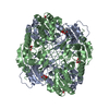 6nxbC  6nxcC  6nxdC C: citing same article ( |
|---|---|
| Similar structure data |

-Links
 PDBj
PDBj

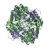
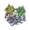
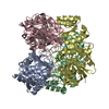
- Assembly
| Deposited unit | 
| ||||||||||||
|---|---|---|---|---|---|---|---|---|---|---|---|---|---|
| 1 | 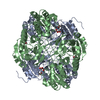
| ||||||||||||
| 2 | 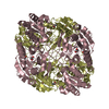
| ||||||||||||
| Unit cell |
| ||||||||||||
| Components on special symmetry positions |
|
-Components
| #1: Protein | Asparaginase / L-asparaginase II / L-ASNase II / L-asparagine amidohydrolase II Mass: 35544.820 Da / Num. of mol.: 4 / Mutation: K162T Source method: isolated from a genetically manipulated source Source: (gene. exp.) Escherichia coli (strain K12) (bacteria)Strain: K12 / Gene: ansB, b2957, JW2924 / Plasmid: pET-22b Details (production host): ORF contains a secretion sequence, 'HHHHHH' affinity tag and sequence of doubly mutated mature EcAII Cell (production host): mesophilic bacteria / Cell line (production host): JC2 / Production host: Escherichia coli BL21(DE3) (bacteria) / Strain (production host): BL21 (DE3) / Variant (production host): ansA, ansB, iaaA triple knockout / References: UniProt: P00805, asparaginase#2: Chemical | ChemComp-ACY / Acetic acid  Mass: 60.052 Da / Num. of mol.: 4 / Source method: obtained synthetically / Formula: C2H4O2 / Feature type: SUBJECT OF INVESTIGATION Mass: 60.052 Da / Num. of mol.: 4 / Source method: obtained synthetically / Formula: C2H4O2 / Feature type: SUBJECT OF INVESTIGATION#3: Chemical | Glycerol  Mass: 92.094 Da / Num. of mol.: 3 / Source method: obtained synthetically / Formula: C3H8O3 / Feature type: SUBJECT OF INVESTIGATION Mass: 92.094 Da / Num. of mol.: 3 / Source method: obtained synthetically / Formula: C3H8O3 / Feature type: SUBJECT OF INVESTIGATION#4: Water | ChemComp-HOH / | Water Mass: 18.015 Da / Num. of mol.: 1181 / Source method: isolated from a natural source / Formula: H2O Mass: 18.015 Da / Num. of mol.: 1181 / Source method: isolated from a natural source / Formula: H2O |
|---|
-Experimental details
-Experiment
| Experiment | Method: X-RAY DIFFRACTION / Number of used crystals: 1 |
|---|
- Sample preparation
| Crystal | Density Matthews: 2.11 Å3/Da / Density % sol: 41.61 % |
|---|---|
| Crystal grow | Temperature: 293 K / Method: vapor diffusion, hanging drop / pH: 7 Details: Protein, at the concentration 15 mg/ml in 50 mM HEPES buffer pH 7 and 150 mM sodium chloride was mixed with equivolume solution of precipitant that contained, 17% (w/v) PEG3350 and 0.17 M ...Details: Protein, at the concentration 15 mg/ml in 50 mM HEPES buffer pH 7 and 150 mM sodium chloride was mixed with equivolume solution of precipitant that contained, 17% (w/v) PEG3350 and 0.17 M ammonium citrate pH 7. Resulting droplets were equilibrated against the precipitant. For the data collection, crystal was briefly transferred to cryo-protecting solution, which had the same composition as precipitant, except concentration of PEG3350 was increased to 35 % (v/w/) and 10% (v/v), and also contained 15% of glycerol Temp details: incubator-controlled |
-Data collection
| Diffraction | Mean temperature: 100 K / Ambient temp details: Stream of liquid nitrogen / Serial crystal experiment: N | |||||||||||||||||||||||||||||||||||||||||||||||||||||||||||||||||||||||||||||||||||||||||||||||||||||||||||||||||||||||||||||||||||||||||||||||||||||||||||||||||||||||||||||||||||||||||||||
|---|---|---|---|---|---|---|---|---|---|---|---|---|---|---|---|---|---|---|---|---|---|---|---|---|---|---|---|---|---|---|---|---|---|---|---|---|---|---|---|---|---|---|---|---|---|---|---|---|---|---|---|---|---|---|---|---|---|---|---|---|---|---|---|---|---|---|---|---|---|---|---|---|---|---|---|---|---|---|---|---|---|---|---|---|---|---|---|---|---|---|---|---|---|---|---|---|---|---|---|---|---|---|---|---|---|---|---|---|---|---|---|---|---|---|---|---|---|---|---|---|---|---|---|---|---|---|---|---|---|---|---|---|---|---|---|---|---|---|---|---|---|---|---|---|---|---|---|---|---|---|---|---|---|---|---|---|---|---|---|---|---|---|---|---|---|---|---|---|---|---|---|---|---|---|---|---|---|---|---|---|---|---|---|---|---|---|---|---|---|---|
| Diffraction source | Source: ROTATING ANODE / Type: RIGAKU MICROMAX-007 HF / Wavelength: 1.5418 Å | |||||||||||||||||||||||||||||||||||||||||||||||||||||||||||||||||||||||||||||||||||||||||||||||||||||||||||||||||||||||||||||||||||||||||||||||||||||||||||||||||||||||||||||||||||||||||||||
| Detector | Type: DECTRIS EIGER R 4M / Detector: PIXEL / Date: Jul 2, 2018 / Details: Multilayer X-ray mirrors VariMax HF | |||||||||||||||||||||||||||||||||||||||||||||||||||||||||||||||||||||||||||||||||||||||||||||||||||||||||||||||||||||||||||||||||||||||||||||||||||||||||||||||||||||||||||||||||||||||||||||
| Radiation | Monochromator: Multilayer X-ray mirrors VariMax HF / Protocol: SINGLE WAVELENGTH / Monochromatic (M) / Laue (L): M / Scattering type: x-ray | |||||||||||||||||||||||||||||||||||||||||||||||||||||||||||||||||||||||||||||||||||||||||||||||||||||||||||||||||||||||||||||||||||||||||||||||||||||||||||||||||||||||||||||||||||||||||||||
| Radiation wavelength | Wavelength: 1.5418 Å / Relative weight: 1 | |||||||||||||||||||||||||||||||||||||||||||||||||||||||||||||||||||||||||||||||||||||||||||||||||||||||||||||||||||||||||||||||||||||||||||||||||||||||||||||||||||||||||||||||||||||||||||||
| Reflection | Resolution: 1.93→40 Å / Num. obs: 86035 / % possible obs: 96.1 % / Redundancy: 3.3 % / Rmerge(I) obs: 0.1 / Rpim(I) all: 0.064 / Rrim(I) all: 0.119 / Χ2: 0.923 / Net I/σ(I): 7.8 / Num. measured all: 284693 | |||||||||||||||||||||||||||||||||||||||||||||||||||||||||||||||||||||||||||||||||||||||||||||||||||||||||||||||||||||||||||||||||||||||||||||||||||||||||||||||||||||||||||||||||||||||||||||
| Reflection shell | Diffraction-ID: 1
|
- Processing
| Software |
| ||||||||||||||||||||||||||||||||||||||||||||||||||||||||||||
|---|---|---|---|---|---|---|---|---|---|---|---|---|---|---|---|---|---|---|---|---|---|---|---|---|---|---|---|---|---|---|---|---|---|---|---|---|---|---|---|---|---|---|---|---|---|---|---|---|---|---|---|---|---|---|---|---|---|---|---|---|---|
| Refinement | Method to determine structure: FOURIER SYNTHESIS / Resolution: 1.93→26.54 Å / Cor.coef. Fo:Fc: 0.963 / Cor.coef. Fo:Fc free: 0.914 / SU B: 4.329 / SU ML: 0.122 / SU R Cruickshank DPI: 0.1547 / Cross valid method: THROUGHOUT / σ(F): 0 / ESU R: 0.155 / ESU R Free: 0.161 Details: HYDROGENS HAVE BEEN ADDED IN THE RIDING POSITIONS U VALUES : REFINED INDIVIDUALLY
| ||||||||||||||||||||||||||||||||||||||||||||||||||||||||||||
| Solvent computation | Ion probe radii: 0.8 Å / Shrinkage radii: 0.8 Å / VDW probe radii: 1.2 Å | ||||||||||||||||||||||||||||||||||||||||||||||||||||||||||||
| Displacement parameters | Biso max: 114.3 Å2 / Biso mean: 33.522 Å2 / Biso min: 15.22 Å2
| ||||||||||||||||||||||||||||||||||||||||||||||||||||||||||||
| Refinement step | Cycle: final / Resolution: 1.93→26.54 Å
| ||||||||||||||||||||||||||||||||||||||||||||||||||||||||||||
| Refine LS restraints |
| ||||||||||||||||||||||||||||||||||||||||||||||||||||||||||||
| LS refinement shell | Resolution: 1.93→1.98 Å / Rfactor Rfree error: 0 / Total num. of bins used: 20
|
