 Movie
Movie Controller

[English] 日本語
 Yorodumi
- PDB-1jja: CRYSTAL STRUCTURE OF ORTHORHOMBIC FORM OF D90E MUTANT OF ESCHERIC... -
+ Open data

- Basic information
| Entry | Database: PDB / ID: 1jja | ||||||
|---|---|---|---|---|---|---|---|
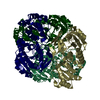
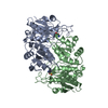
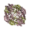
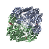
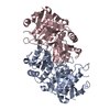
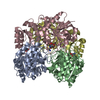
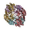
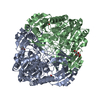
| Title | CRYSTAL STRUCTURE OF ORTHORHOMBIC FORM OF D90E MUTANT OF ESCHERICHIA COLI L-ASPARAGINASE II | ||||||
 Components Components | L-ASPARAGINASE II Asparaginase Asparaginase | ||||||
 Keywords Keywords | HYDROLASE / L-ASPARAGINASE / LEUKEMIA | ||||||
| Function / homology |  Function and homology information Function and homology informationasparagine catabolic process / asparaginase / asparaginase activity / outer membrane-bounded periplasmic space / protein homotetramerization / periplasmic space / protein-containing complex / identical protein bindingSimilarity search - Function | ||||||
| Biological species |  Escherichia coli (E. coli) Escherichia coli (E. coli) | ||||||
| Method | X-RAY DIFFRACTION / MOLECULAR REPLACEMENT / Resolution: 2.3 Å | ||||||
 Authors Authors | Borek, D. / Kozak, M. / Jaskolski, M. | ||||||
 Citation Citation | Journal: Febs J. / Year: 2014 Title: Crystal structure of active site mutant of antileukemic L-asparaginase reveals conserved zinc-binding site. Authors: Borek, D. / Kozak, M. / Pei, J. / Jaskolski, M. #1: Journal: Proc.Natl.Acad.Sci.USA / Year: 1993Title: Crystal Structure of Escherichia coli L-asparaginase, An Enzyme Used in Cancer Therapy Authors: Swain, A.L. / Jaskolski, M. / Housset, D. / Rao, J.K. / Wlodawer, A. #2: Journal: FEBS Lett. / Year: 1996Title: A Covalently Bound Catalytic Intermediate in Escherichia coli Asparaginase: Crystal Structure of a Thr-89-Val Mutant Authors: Palm, G.J. / Lubkowski, J. / Derst, C. / Schleper, S. / Rohm, K.H. / Wlodawer, A. #3: Journal: Acta Crystallogr.,Sect.D / Year: 2001Title: Structures of Two Highly Homologous Bacterial L-Asparaginases: A Case of Enantiomorphic Space Groups Authors: Jaskolski, M. / Kozak, M. / Lubkowski, J. / Palm, G. / Wlodawer, A. #4: Journal: ACTA BIOCHIM.POL. / Year: 1997Title: Why a "benign" Mutation Kills Enzyme Activity. Structure-based Analysis of the A176V Mutant of Saccharomyces cerevisiae L-asparaginase I Authors: Bonthron, D.T. / Jaskolski, M. #5: Journal: BIOCHIM.BIOPHYS.ACTA / Year: 2000Title: Dynamics of a Mobile Loop at the Active Site of Escherichia coli Asparaginase Authors: Aung, H.P. / Bocola, M. / Schleper, S. / Rohm, K.H. | ||||||
| History |
| ||||||
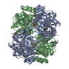
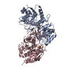
| Remark 300 | BIOMOLECULE: 1, 2 THIS ENTRY CONTAINS THE CRYSTALLOGRAPHIC ASYMMETRIC UNIT WHICH CONSISTS OF 6 ...BIOMOLECULE: 1, 2 THIS ENTRY CONTAINS THE CRYSTALLOGRAPHIC ASYMMETRIC UNIT WHICH CONSISTS OF 6 CHAINS, A, B, C, D, E, F. THE BIOLOGICAL ASSEMBLY IS A HOMOTETRAMER. CHAINS A, B, C, D FORM ONE INDEPENDENT TETRAMER WITH NON-CRYSTALLOGRAPHIC 222 SYMMETRY. CHAINS E AND F FORM AN ACTIVE-SITE-COMPETENT DIMER (CORRESPONDING TO DIMER AC IN THE ABCD TETRAMER). THE COMPLETE EFE'F' TETRAMER IS GENERATED THROUGH THE CRYSTALLOGRAPHIC TWO-FOLD ROTATION. SEE REMARK 350 FOR INFORMATION ON GENERATING THE BIOLOGICAL MOLECULE(S). |
- Structure visualization
| Structure viewer | Molecule: MolmilJmol/JSmol |
|---|
- Downloads & links
-Download
| PDBx/mmCIF format | 1jja.cif.gz | 355.7 KB | Display | PDBx/mmCIF format |
|---|---|---|---|---|
| PDB format | pdb1jja.ent.gz | 291.9 KB | Display | PDB format |
| PDBx/mmJSON format | 1jja.json.gz | Tree view | PDBx/mmJSON format | |
| Others |  Other downloads Other downloads |
-Validation report
| Arichive directory | https://data.pdbj.org/pub/pdb/validation_reports/jj/1jjaftp://data.pdbj.org/pub/pdb/validation_reports/jj/1jja | HTTPS FTP |
|---|
-Related structure data
| Related structure data |  1ihdC 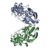 1jazC  3ecaS C: citing same article ( S: Starting model for refinement |
|---|---|
| Similar structure data |


-Links
 PDBj
PDBj- Assembly
| Deposited unit | 
| ||||||||
|---|---|---|---|---|---|---|---|---|---|
| 1 | 
| ||||||||
| 2 | 
| ||||||||
| Unit cell |
| ||||||||
| Details | The biological assembly is a homotetramer. The asymmetric unit contains six protein chains, A, B, C, D, E, F. Chains A, B, C, D form one independent tetramer with non-crystallographic 222 symmetry. Chains E and F form an active-site-competent dimer (corresponding to dimer AC in the ABCD tetramer). The complete EFE'F' tetramer is generated through the crystallographic two-fold rotation. |
-Components
| #1: Protein | Asparaginase / L-ASNASE II Mass: 34640.836 Da / Num. of mol.: 6 / Mutation: D90E Source method: isolated from a genetically manipulated source Source: (gene. exp.) Escherichia coli (E. coli) / Production host: Escherichia coli (E. coli) / References: UniProt: P00805, asparaginase#2: Water | ChemComp-HOH / | Water Mass: 18.015 Da / Num. of mol.: 444 / Source method: isolated from a natural source / Formula: H2O Mass: 18.015 Da / Num. of mol.: 444 / Source method: isolated from a natural source / Formula: H2O |
|---|
-Experimental details
-Experiment
| Experiment | Method: X-RAY DIFFRACTION / Number of used crystals: 1 |
|---|
- Sample preparation
| Crystal | Density Matthews: 2.17 Å3/Da / Density % sol: 43 % |
|---|---|
| Crystal grow | Temperature: 292 K / Method: vapor diffusion, hanging drop / pH: 9 Details: PEG MME550, bicine, NaCl , pH 9.0, VAPOR DIFFUSION, HANGING DROP, temperature 292K |
-Data collection
| Diffraction | Mean temperature: 290 K |
|---|---|
| Diffraction source | Source: ROTATING ANODE / Type: SIEMENS / Wavelength: 1.54178 Å |
| Detector | Type: MARRESEARCH / Detector: IMAGE PLATE / Date: Sep 10, 1998 |
| Radiation | Monochromator: GRAPHITE / Protocol: SINGLE WAVELENGTH / Monochromatic (M) / Laue (L): M / Scattering type: x-ray |
| Radiation wavelength | Wavelength: 1.54178 Å / Relative weight: 1 |
| Reflection | Resolution: 2.3→25 Å / Num. all: 75961 / Num. obs: 75961 / Observed criterion σ(F): 0 / Observed criterion σ(I): -3 / Redundancy: 3.14 % / Rmerge(I) obs: 0.123 / Net I/σ(I): 11.2 |
| Reflection shell | Resolution: 2.3→2.41 Å / Redundancy: 3.07 % / Rmerge(I) obs: 0.813 / Mean I/σ(I) obs: 1.8 / % possible all: 93.9 |
- Processing
| Software |
| ||||||||||||||||||||||||||||||||||||||||||||||||||||||||||||||||||||||||||||||||||||
|---|---|---|---|---|---|---|---|---|---|---|---|---|---|---|---|---|---|---|---|---|---|---|---|---|---|---|---|---|---|---|---|---|---|---|---|---|---|---|---|---|---|---|---|---|---|---|---|---|---|---|---|---|---|---|---|---|---|---|---|---|---|---|---|---|---|---|---|---|---|---|---|---|---|---|---|---|---|---|---|---|---|---|---|---|---|
| Refinement | Method to determine structure: MOLECULAR REPLACEMENT Starting model: Active dimer of native E.coli asparaginase II (PDB code: 3ECA) Resolution: 2.3→10 Å / SU B: 5.781 / SU ML: 0.136 / Isotropic thermal model: ISOTROPIC / Cross valid method: THROUGHOUT / σ(F): 0 / ESU R: 0.40726 / ESU R Free: 0 Details: MAXIMUM LIKELIHOOD METHOD. RESIDUES 17-33 IN MONOMER A, 16-37 IN MONOMER B, 16-34 IN MONOMER D, 16-34 IN MONOMER E, 19-20 AND 30-33 IN MONOMER F ARE NOT PRESENT IN THE MODEL. IN MONOMER C, ...Details: MAXIMUM LIKELIHOOD METHOD. RESIDUES 17-33 IN MONOMER A, 16-37 IN MONOMER B, 16-34 IN MONOMER D, 16-34 IN MONOMER E, 19-20 AND 30-33 IN MONOMER F ARE NOT PRESENT IN THE MODEL. IN MONOMER C, THE COMPLETE FLEXIBLE LOOP BETWEEN RESIDUES 10 AND 40 HAS BEEN MODELED IN VERY GOOD ELECTRON DENSITY IN AN OPEN CONFORMATION.
| ||||||||||||||||||||||||||||||||||||||||||||||||||||||||||||||||||||||||||||||||||||
| Displacement parameters | Biso mean: 39.399 Å2
| ||||||||||||||||||||||||||||||||||||||||||||||||||||||||||||||||||||||||||||||||||||
| Refinement step | Cycle: LAST / Resolution: 2.3→10 Å
| ||||||||||||||||||||||||||||||||||||||||||||||||||||||||||||||||||||||||||||||||||||
| Refine LS restraints |
|
