 Movie
Movie Controller


+ Open data

- Basic information
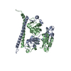
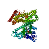
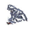
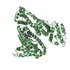
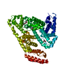
| Entry | Database: PDB / ID: 6ncq | ||||||
|---|---|---|---|---|---|---|---|
| Title | The dimerization domain of human SFPQ in space group C2221 | ||||||
 Components Components | Splicing factor, proline- and glutamine-rich | ||||||
 Keywords Keywords | NUCLEAR PROTEIN / DBHS protein RNA binding protein Gene regulation | ||||||
| Function / homology |  Function and homology information Function and homology informationPTK6 Regulates Proteins Involved in RNA Processing / negative regulation of circadian rhythm / alternative mRNA splicing, via spliceosome / Suppression of apoptosis / paraspeckles / positive regulation of oxidative stress-induced intrinsic apoptotic signaling pathway / activation of innate immune response / RNA splicing / double-strand break repair via homologous recombination / regulation of circadian rhythm ...PTK6 Regulates Proteins Involved in RNA Processing / negative regulation of circadian rhythm / alternative mRNA splicing, via spliceosome / Suppression of apoptosis / paraspeckles / positive regulation of oxidative stress-induced intrinsic apoptotic signaling pathway / activation of innate immune response / RNA splicing / double-strand break repair via homologous recombination / regulation of circadian rhythm / mRNA processing / nuclear matrix / histone deacetylase binding / RNA polymerase II transcription regulator complex / rhythmic process / transcription cis-regulatory region binding / nuclear speck / chromatin remodeling / innate immune response / negative regulation of DNA-templated transcription / chromatin binding / chromatin / regulation of DNA-templated transcription / negative regulation of transcription by RNA polymerase II / protein homodimerization activity / positive regulation of transcription by RNA polymerase II / DNA binding / RNA binding / nucleoplasm / nucleus / cytosolSimilarity search - Function | ||||||
| Biological species |  Homo sapiens (human) Homo sapiens (human) | ||||||
| Method | X-RAY DIFFRACTION / SYNCHROTRON / MOLECULAR REPLACEMENT / Resolution: 1.9 Å | ||||||
 Authors Authors | Lee, M. | ||||||
| Funding support |  Australia, 1items Australia, 1items
| ||||||
 Citation Citation | Journal: Acta Crystallogr.,Sect.F / Year: 2019 Title: A new crystal structure and small-angle X-ray scattering analysis of the homodimer of human SFPQ. Authors: Hewage, T.W. / Caria, S. / Lee, M. | ||||||
| History |
|
- Structure visualization
| Structure viewer | Molecule: MolmilJmol/JSmol |
|---|
- Downloads & links
-Download
| PDBx/mmCIF format | 6ncq.cif.gz | 72.1 KB | Display | PDBx/mmCIF format |
|---|---|---|---|---|
| PDB format | pdb6ncq.ent.gz | 51.2 KB | Display | PDB format |
| PDBx/mmJSON format | 6ncq.json.gz | Tree view | PDBx/mmJSON format | |
| Others |  Other downloads Other downloads |
-Validation report
| Arichive directory | https://data.pdbj.org/pub/pdb/validation_reports/nc/6ncqftp://data.pdbj.org/pub/pdb/validation_reports/nc/6ncq | HTTPS FTP |
|---|
-Related structure data
| Related structure data |  4wiiS S: Starting model for refinement |
|---|---|
| Similar structure data |


-Links
 PDBj
PDBj- Assembly
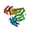
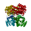
| Deposited unit | 
| ||||||||
|---|---|---|---|---|---|---|---|---|---|
| 1 | 
| ||||||||
| Unit cell |
|
-Components
| #1: Protein | / 100 kDa DNA-pairing protein / hPOMp100 / DNA-binding p52/p100 complex / 100 kDa subunit / ...100 kDa DNA-pairing protein / hPOMp100 / DNA-binding p52/p100 complex / 100 kDa subunit / Polypyrimidine tract-binding protein-associated-splicing factor / PTB-associated-splicing factor Mass: 30094.045 Da / Num. of mol.: 1 Source method: isolated from a genetically manipulated source Source: (gene. exp.) Homo sapiens (human) / Gene: SFPQ, PSF / Production host:  Escherichia coli (E. coli) / References: UniProt: P23246 Escherichia coli (E. coli) / References: UniProt: P23246 |
|---|---|
| #2: Water | ChemComp-HOH / Water Mass: 18.015 Da / Num. of mol.: 208 / Source method: isolated from a natural source / Formula: H2O Mass: 18.015 Da / Num. of mol.: 208 / Source method: isolated from a natural source / Formula: H2O |
-Experimental details
-Experiment
| Experiment | Method: X-RAY DIFFRACTION / Number of used crystals: 1 |
|---|
- Sample preparation
| Crystal | Density Matthews: 2.35 Å3/Da / Density % sol: 47.57 % |
|---|---|
| Crystal grow | Temperature: 293 K / Method: vapor diffusion, hanging drop / Details: 0.1 M Tris-HCl (pH 8,5), 35-40% (v/v) MPD |
-Data collection
| Diffraction | Mean temperature: 100 K / Serial crystal experiment: N |
|---|---|
| Diffraction source | Source: SYNCHROTRON / Site: Australian Synchrotron / Beamline: MX2 / Wavelength: 0.9537 Å |
| Detector | Type: ADSC QUANTUM 315r / Detector: CCD / Date: Apr 7, 2016 |
| Radiation | Protocol: SINGLE WAVELENGTH / Monochromatic (M) / Laue (L): M / Scattering type: x-ray |
| Radiation wavelength | Wavelength: 0.9537 Å / Relative weight: 1 |
| Reflection | Resolution: 1.9→20.49 Å / Num. obs: 22646 / % possible obs: 99.8 % / Redundancy: 4 % / Biso Wilson estimate: 30.32 Å2 / CC1/2: 0.999 / Net I/σ(I): 15.8 |
| Reflection shell | Resolution: 1.9→1.95 Å / Redundancy: 4.1 % / Num. unique obs: 1436 / CC1/2: 0.827 / % possible all: 100 |
- Processing
| Software |
| ||||||||||||||||||||||||||||||||||||||||||||||||||||||||||||||||||||||||||||||||||||||||||||||||||||||||||||||||||
|---|---|---|---|---|---|---|---|---|---|---|---|---|---|---|---|---|---|---|---|---|---|---|---|---|---|---|---|---|---|---|---|---|---|---|---|---|---|---|---|---|---|---|---|---|---|---|---|---|---|---|---|---|---|---|---|---|---|---|---|---|---|---|---|---|---|---|---|---|---|---|---|---|---|---|---|---|---|---|---|---|---|---|---|---|---|---|---|---|---|---|---|---|---|---|---|---|---|---|---|---|---|---|---|---|---|---|---|---|---|---|---|---|---|---|---|
| Refinement | Method to determine structure: MOLECULAR REPLACEMENT Starting model: 4wii Resolution: 1.9→20.49 Å / Cor.coef. Fo:Fc: 0.93 / Cor.coef. Fo:Fc free: 0.912 / SU R Cruickshank DPI: 0.159 / Cross valid method: THROUGHOUT / σ(F): 0 / SU R Blow DPI: 0.17 / SU Rfree Blow DPI: 0.152 / SU Rfree Cruickshank DPI: 0.147
| ||||||||||||||||||||||||||||||||||||||||||||||||||||||||||||||||||||||||||||||||||||||||||||||||||||||||||||||||||
| Displacement parameters | Biso mean: 37.33 Å2
| ||||||||||||||||||||||||||||||||||||||||||||||||||||||||||||||||||||||||||||||||||||||||||||||||||||||||||||||||||
| Refine analyze | Luzzati coordinate error obs: 0.28 Å | ||||||||||||||||||||||||||||||||||||||||||||||||||||||||||||||||||||||||||||||||||||||||||||||||||||||||||||||||||
| Refinement step | Cycle: 1 / Resolution: 1.9→20.49 Å
| ||||||||||||||||||||||||||||||||||||||||||||||||||||||||||||||||||||||||||||||||||||||||||||||||||||||||||||||||||
| Refine LS restraints |
| ||||||||||||||||||||||||||||||||||||||||||||||||||||||||||||||||||||||||||||||||||||||||||||||||||||||||||||||||||
| LS refinement shell | Resolution: 1.9→1.91 Å / Total num. of bins used: 50
|
