 Movie
Movie Controller


+ Open data

- Basic information
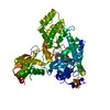
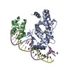
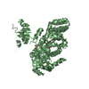
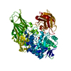
| Entry | Database: PDB / ID: 4wii | |||||||||||||||
|---|---|---|---|---|---|---|---|---|---|---|---|---|---|---|---|---|
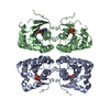
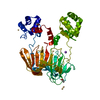
| Title | HUMAN SPLICING FACTOR, CONSTRUCT 3 | |||||||||||||||
 Components Components | Splicing factor, proline- and glutamine-rich | |||||||||||||||
 Keywords Keywords | TRANSCRIPTION / RRM | |||||||||||||||
| Function / homology |  Function and homology information Function and homology informationPTK6 Regulates Proteins Involved in RNA Processing / negative regulation of circadian rhythm / alternative mRNA splicing, via spliceosome / Suppression of apoptosis / paraspeckles / positive regulation of oxidative stress-induced intrinsic apoptotic signaling pathway / activation of innate immune response / RNA splicing / double-strand break repair via homologous recombination / regulation of circadian rhythm ...PTK6 Regulates Proteins Involved in RNA Processing / negative regulation of circadian rhythm / alternative mRNA splicing, via spliceosome / Suppression of apoptosis / paraspeckles / positive regulation of oxidative stress-induced intrinsic apoptotic signaling pathway / activation of innate immune response / RNA splicing / double-strand break repair via homologous recombination / regulation of circadian rhythm / mRNA processing / nuclear matrix / histone deacetylase binding / RNA polymerase II transcription regulator complex / rhythmic process / transcription cis-regulatory region binding / nuclear speck / chromatin remodeling / innate immune response / negative regulation of DNA-templated transcription / chromatin binding / chromatin / regulation of DNA-templated transcription / negative regulation of transcription by RNA polymerase II / protein homodimerization activity / positive regulation of transcription by RNA polymerase II / DNA binding / RNA binding / nucleoplasm / nucleus / cytosolSimilarity search - Function | |||||||||||||||
| Biological species |  Homo sapiens (human) Homo sapiens (human) | |||||||||||||||
| Method | X-RAY DIFFRACTION / SYNCHROTRON / MOLECULAR REPLACEMENT / Resolution: 2.05 Å | |||||||||||||||
 Authors Authors | lee, M. / bond, c.s. | |||||||||||||||
| Funding support |  Australia, 4items Australia, 4items
| |||||||||||||||
 Citation Citation | Journal: Nucleic Acids Res. / Year: 2015 Title: The structure of human SFPQ reveals a coiled-coil mediated polymer essential for functional aggregation in gene regulation. Authors: Lee, M. / Sadowska, A. / Bekere, I. / Ho, D. / Gully, B.S. / Lu, Y. / Iyer, K.S. / Trewhella, J. / Fox, A.H. / Bond, C.S. | |||||||||||||||
| History |
|
- Structure visualization
| Structure viewer | Molecule: MolmilJmol/JSmol |
|---|
- Downloads & links
-Download
| PDBx/mmCIF format | 4wii.cif.gz | 117.6 KB | Display | PDBx/mmCIF format |
|---|---|---|---|---|
| PDB format | pdb4wii.ent.gz | 89.6 KB | Display | PDB format |
| PDBx/mmJSON format | 4wii.json.gz | Tree view | PDBx/mmJSON format | |
| Others |  Other downloads Other downloads |
-Validation report
| Arichive directory | https://data.pdbj.org/pub/pdb/validation_reports/wi/4wiiftp://data.pdbj.org/pub/pdb/validation_reports/wi/4wii | HTTPS FTP |
|---|
-Related structure data


-Links
 PDBj
PDBj- Assembly
| Deposited unit | 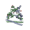
| ||||||||
|---|---|---|---|---|---|---|---|---|---|
| 1 |
| ||||||||
| Unit cell |
|
-Components
| #1: Protein | / 100 kDa DNA-pairing protein / hPOMp100 / DNA-binding p52/p100 complex / 100 kDa subunit / ...100 kDa DNA-pairing protein / hPOMp100 / DNA-binding p52/p100 complex / 100 kDa subunit / Polypyrimidine tract-binding protein-associated-splicing factor / PTB-associated-splicing factor Mass: 30094.045 Da / Num. of mol.: 2 / Fragment: UNP RESIDUES 276-535 Source method: isolated from a genetically manipulated source Source: (gene. exp.) Homo sapiens (human) / Gene: SFPQ, PSF / Production host:  Escherichia coli (E. coli) / References: UniProt: P23246 Escherichia coli (E. coli) / References: UniProt: P23246#2: Chemical | ChemComp-SO4 / | Sulfate  Mass: 96.063 Da / Num. of mol.: 1 / Source method: obtained synthetically / Formula: SO4 Mass: 96.063 Da / Num. of mol.: 1 / Source method: obtained synthetically / Formula: SO4#3: Chemical | ChemComp-EDO / Ethylene glycol  Mass: 62.068 Da / Num. of mol.: 8 / Source method: obtained synthetically / Formula: C2H6O2 Mass: 62.068 Da / Num. of mol.: 8 / Source method: obtained synthetically / Formula: C2H6O2#4: Water | ChemComp-HOH / | Water Mass: 18.015 Da / Num. of mol.: 176 / Source method: isolated from a natural source / Formula: H2O Mass: 18.015 Da / Num. of mol.: 176 / Source method: isolated from a natural source / Formula: H2O |
|---|
-Experimental details
-Experiment
| Experiment | Method: X-RAY DIFFRACTION / Number of used crystals: 1 |
|---|
- Sample preparation
| Crystal | Density Matthews: 2.13 Å3/Da / Density % sol: 42.25 % |
|---|---|
| Crystal grow | Temperature: 293 K / Method: vapor diffusion, hanging drop / pH: 8.5 Details: 1.9 M AMMONIUM SULFATE, 0.1 M TRIS, 20% ETHYLENE GLYCOL (V/V), PH 8.5, VAPOR DIFFUSION, HANGING DROP, TEMPERATURE 293K PH range: 8.5 |
-Data collection
| Diffraction | Mean temperature: 100 K |
|---|---|
| Diffraction source | Source: SYNCHROTRON / Site: Australian Synchrotron / Beamline: MX1 / Wavelength: 0.9686 Å |
| Detector | Type: ADSC QUANTUM 210r / Detector: CCD / Date: Oct 15, 2010 |
| Radiation | Monochromator: DIPOLE/BENDING MAGNET / Protocol: SINGLE WAVELENGTH / Monochromatic (M) / Laue (L): M / Scattering type: x-ray |
| Radiation wavelength | Wavelength: 0.9686 Å / Relative weight: 1 |
| Reflection | Resolution: 2.05→19.61 Å / Num. obs: 32938 / % possible obs: 99.9 % / Observed criterion σ(I): 0 / Redundancy: 7.1 % / Biso Wilson estimate: 36.61 Å2 / Net I/σ(I): 13.1 |
| Reflection shell | Resolution: 2.05→2.16 Å / Mean I/σ(I) obs: 2.1 / % possible all: 100 |
- Processing
| Software |
| ||||||||||||||||||||||||||||||||||||||||||||||||||||||||||||||||||||||||||||||||||||||||||||||||||||||||||||||||||
|---|---|---|---|---|---|---|---|---|---|---|---|---|---|---|---|---|---|---|---|---|---|---|---|---|---|---|---|---|---|---|---|---|---|---|---|---|---|---|---|---|---|---|---|---|---|---|---|---|---|---|---|---|---|---|---|---|---|---|---|---|---|---|---|---|---|---|---|---|---|---|---|---|---|---|---|---|---|---|---|---|---|---|---|---|---|---|---|---|---|---|---|---|---|---|---|---|---|---|---|---|---|---|---|---|---|---|---|---|---|---|---|---|---|---|---|
| Refinement | Method to determine structure: MOLECULAR REPLACEMENT / Resolution: 2.05→19.52 Å / Cor.coef. Fo:Fc: 0.934 / Cor.coef. Fo:Fc free: 0.921 / SU R Cruickshank DPI: 0.21 / Cross valid method: THROUGHOUT / σ(F): 0
| ||||||||||||||||||||||||||||||||||||||||||||||||||||||||||||||||||||||||||||||||||||||||||||||||||||||||||||||||||
| Displacement parameters | Biso mean: 43.79 Å2
| ||||||||||||||||||||||||||||||||||||||||||||||||||||||||||||||||||||||||||||||||||||||||||||||||||||||||||||||||||
| Refine analyze | Luzzati coordinate error obs: 0.27 Å | ||||||||||||||||||||||||||||||||||||||||||||||||||||||||||||||||||||||||||||||||||||||||||||||||||||||||||||||||||
| Refinement step | Cycle: 1 / Resolution: 2.05→19.52 Å
| ||||||||||||||||||||||||||||||||||||||||||||||||||||||||||||||||||||||||||||||||||||||||||||||||||||||||||||||||||
| Refine LS restraints |
| ||||||||||||||||||||||||||||||||||||||||||||||||||||||||||||||||||||||||||||||||||||||||||||||||||||||||||||||||||
| LS refinement shell | Resolution: 2.05→2.12 Å / Total num. of bins used: 16
|
