 Movie
Movie Controller


+ Open data

- Basic information
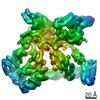
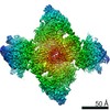
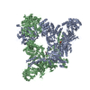
| Entry | Database: PDB / ID: 6lxv | ||||||
|---|---|---|---|---|---|---|---|
| Title | Cryo-EM structure of phosphoketolase from Bifidobacterium longum | ||||||
 Components Components | Phosphoketolase | ||||||
 Keywords Keywords | LYASE / ketolase / thiamine diphosphate / octamer / Bifidobacterium longum / lyase activity | ||||||
| Function / homology |  Function and homology informationfructose-6-phosphate phosphoketolase / fructose-6-phosphate phosphoketolase activity / carbohydrate metabolic process Function and homology informationfructose-6-phosphate phosphoketolase / fructose-6-phosphate phosphoketolase activity / carbohydrate metabolic processSimilarity search - Function | ||||||
| Biological species |  Bifidobacterium longum subsp. longum F8 (bacteria) Bifidobacterium longum subsp. longum F8 (bacteria) | ||||||
| Method | ELECTRON MICROSCOPY / single particle reconstruction / cryo EM / Resolution: 2.1 Å | ||||||
 Authors Authors | Nakata, K. / Miyazaki, N. / Yamaguchi, H. / Hirose, M. / Miyano, H. / Mizukoshi, T. / Kashiwagi, T. / Iwasaki, K. | ||||||
| Funding support |  Japan, 1items Japan, 1items
| ||||||
 Citation Citation | Journal: J Struct Biol / Year: 2022 Title: High-resolution structure of phosphoketolase from Bifidobacterium longum determined by cryo-EM single-particle analysis. Authors: Kunio Nakata / Naoyuki Miyazaki / Hiroki Yamaguchi / Mika Hirose / Tatsuki Kashiwagi / Nidamarthi H V Kutumbarao / Osamu Miyashita / Florence Tama / Hiroshi Miyano / Toshimi Mizukoshi / Kenji Iwasaki / Abstract: In bifidobacteria, phosphoketolase (PKT) plays a key role in the central hexose fermentation pathway called "bifid shunt." The three-dimensional structure of PKT from Bifidobacterium longum with co- ...In bifidobacteria, phosphoketolase (PKT) plays a key role in the central hexose fermentation pathway called "bifid shunt." The three-dimensional structure of PKT from Bifidobacterium longum with co-enzyme thiamine diphosphate (ThDpp) was determined at 2.1 Å resolution by cryo-EM single-particle analysis using 196,147 particles to build up the structural model of a PKT octamer related by D symmetry. Although the cryo-EM structure of PKT was almost identical to the X-ray crystal structure previously determined at 2.2 Å resolution, several interesting structural features were observed in the cryo-EM structure. Because this structure was solved at relatively high resolution, it was observed that several amino acid residues adopt multiple conformations. Among them, Q546-D547-H548-N549 (the QN-loop) demonstrate the largest structural change, which seems to be related to the enzymatic function of PKT. The QN-loop is at the entrance to the substrate binding pocket. The minor conformer of the QN-loop is similar to the conformation of the QN-loop in the crystal structure. The major conformer is located further from ThDpp than the minor conformer. Interestingly, the major conformer in the cryo-EM structure of PKT resembles the corresponding loop structure of substrate-bound Escherichia coli transketolase. That is, the minor and major conformers may correspond to "closed" and "open" states for substrate access, respectively. Moreover, because of the high-resolution analysis, many water molecules were observed in the cryo-EM structure of PKT. Structural features of the water molecules in the cryo-EM structure are discussed and compared with water molecules observed in the crystal structure. | ||||||
| History |
|
- Structure visualization
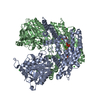
| Movie |
Movie viewer |
|---|---|
| Structure viewer | Molecule: MolmilJmol/JSmol |
- Downloads & links
-Download
| PDBx/mmCIF format | 6lxv.cif.gz | 1.1 MB | Display | PDBx/mmCIF format |
|---|---|---|---|---|
| PDB format | pdb6lxv.ent.gz | 968.3 KB | Display | PDB format |
| PDBx/mmJSON format | 6lxv.json.gz | Tree view | PDBx/mmJSON format | |
| Others |  Other downloads Other downloads |
-Validation report
| Arichive directory | https://data.pdbj.org/pub/pdb/validation_reports/lx/6lxvftp://data.pdbj.org/pub/pdb/validation_reports/lx/6lxv | HTTPS FTP |
|---|
-Related structure data
| Related structure data | 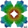 30007MC M: map data used to model this data C: citing same article ( |
|---|---|
| Similar structure data |

-Links
 PDBj
PDBj


- Assembly
| Deposited unit | 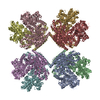
|
|---|---|
| 1 |
|
-Components
| #1: Protein | Mass: 93450.016 Da / Num. of mol.: 8 Source method: isolated from a genetically manipulated source Source: (gene. exp.) Bifidobacterium longum subsp. longum F8 (bacteria)Gene: BIL_11880 / Plasmid: pET24 / Production host: Escherichia coli BL21(DE3) (bacteria)References: UniProt: D6D942, fructose-6-phosphate phosphoketolase#2: Chemical | ChemComp-TPP / Thiamine pyrophosphate  Mass: 425.314 Da / Num. of mol.: 8 / Source method: obtained synthetically / Formula: C12H19N4O7P2S / Feature type: SUBJECT OF INVESTIGATION Mass: 425.314 Da / Num. of mol.: 8 / Source method: obtained synthetically / Formula: C12H19N4O7P2S / Feature type: SUBJECT OF INVESTIGATION#3: Chemical | ChemComp-CA /   Mass: 40.078 Da / Num. of mol.: 8 / Source method: obtained synthetically / Formula: Ca / Feature type: SUBJECT OF INVESTIGATION Mass: 40.078 Da / Num. of mol.: 8 / Source method: obtained synthetically / Formula: Ca / Feature type: SUBJECT OF INVESTIGATION#4: Water | ChemComp-HOH / | Water Mass: 18.015 Da / Num. of mol.: 2967 / Source method: isolated from a natural source / Formula: H2O Mass: 18.015 Da / Num. of mol.: 2967 / Source method: isolated from a natural source / Formula: H2OHas ligand of interest | Y | |
|---|
-Experimental details
-Experiment
| Experiment | Method: ELECTRON MICROSCOPY |
|---|---|
| EM experiment | Aggregation state: PARTICLE / 3D reconstruction method: single particle reconstruction |
- Sample preparation
| Component | Name: Phosphoketolase with thiamine-diphophate / Type: COMPLEX / Entity ID: #1 / Source: RECOMBINANT |
|---|---|
| Molecular weight | Experimental value: NO |
| Source (natural) | Organism: Bifidobacterium longum subsp. longum F8 (bacteria) |
| Source (recombinant) | Organism: Escherichia coli BL21(DE3) (bacteria) / Plasmid: pET24 |
| Buffer solution | pH: 9 |
| Specimen | Conc.: 10 mg/ml / Embedding applied: NO / Shadowing applied: NO / Staining applied: NO / Vitrification applied: YES |
| Specimen support | Grid material: MOLYBDENUM / Grid mesh size: 300 divisions/in. / Grid type: Quantifoil R1.2/1.3 |
| Vitrification | Cryogen name: ETHANE |
- Electron microscopy imaging
| Experimental equipment |  Model: Titan Krios / Image courtesy: FEI Company |
|---|---|
| Microscopy | Model: FEI TITAN KRIOS |
| Electron gun | Electron source: FIELD EMISSION GUN / Accelerating voltage: 300 kV / Illumination mode: FLOOD BEAM |
| Electron lens | Mode: BRIGHT FIELDBright-field microscopy / Nominal magnification: 75000 X / Nominal defocus max: 2750 nm / Nominal defocus min: 1000 nm / Alignment procedure: ZEMLIN TABLEAU |
| Specimen holder | Cryogen: NITROGEN / Specimen holder model: FEI TITAN KRIOS AUTOGRID HOLDER |
| Image recording | Average exposure time: 45 sec. / Electron dose: 50 e/Å2 / Detector mode: COUNTING / Film or detector model: FEI FALCON III (4k x 4k) / Num. of real images: 2897 |
| Image scans | Width: 4096 / Height: 4096 |
- Processing
| EM software |
| ||||||||||||||||||||||||||||||||
|---|---|---|---|---|---|---|---|---|---|---|---|---|---|---|---|---|---|---|---|---|---|---|---|---|---|---|---|---|---|---|---|---|---|
| CTF correction | Type: PHASE FLIPPING AND AMPLITUDE CORRECTION | ||||||||||||||||||||||||||||||||
| Particle selection | Num. of particles selected: 426149 | ||||||||||||||||||||||||||||||||
| Symmetry | Point symmetry: D4 (2x4 fold dihedral) | ||||||||||||||||||||||||||||||||
| 3D reconstruction | Resolution: 2.1 Å / Resolution method: FSC 0.143 CUT-OFF / Num. of particles: 194517 / Algorithm: FOURIER SPACE / Symmetry type: POINT |
